Trademarked Brain Vision blue icon representing EEG

EEG

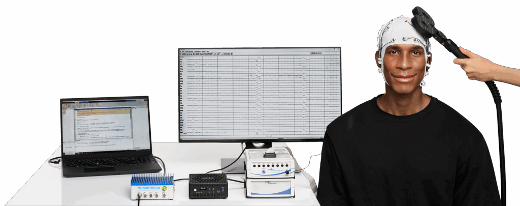
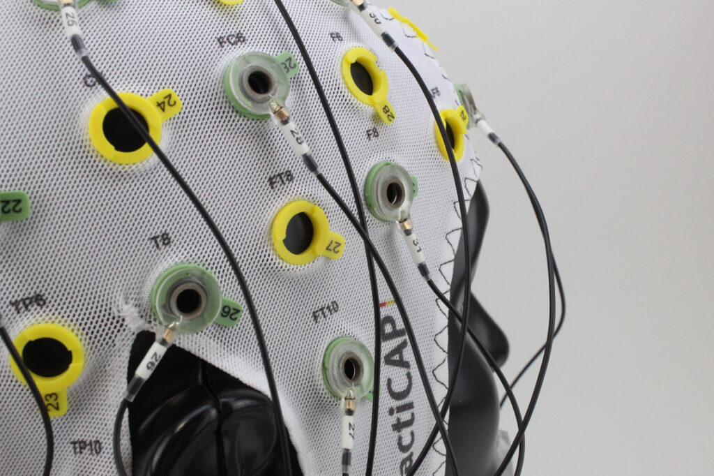
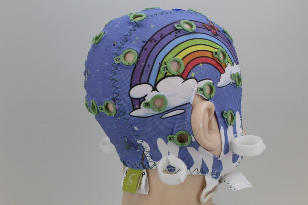
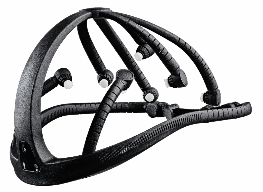
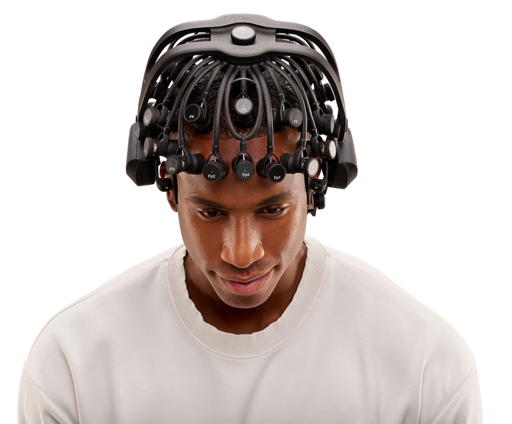
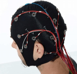
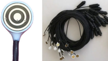
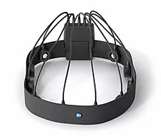
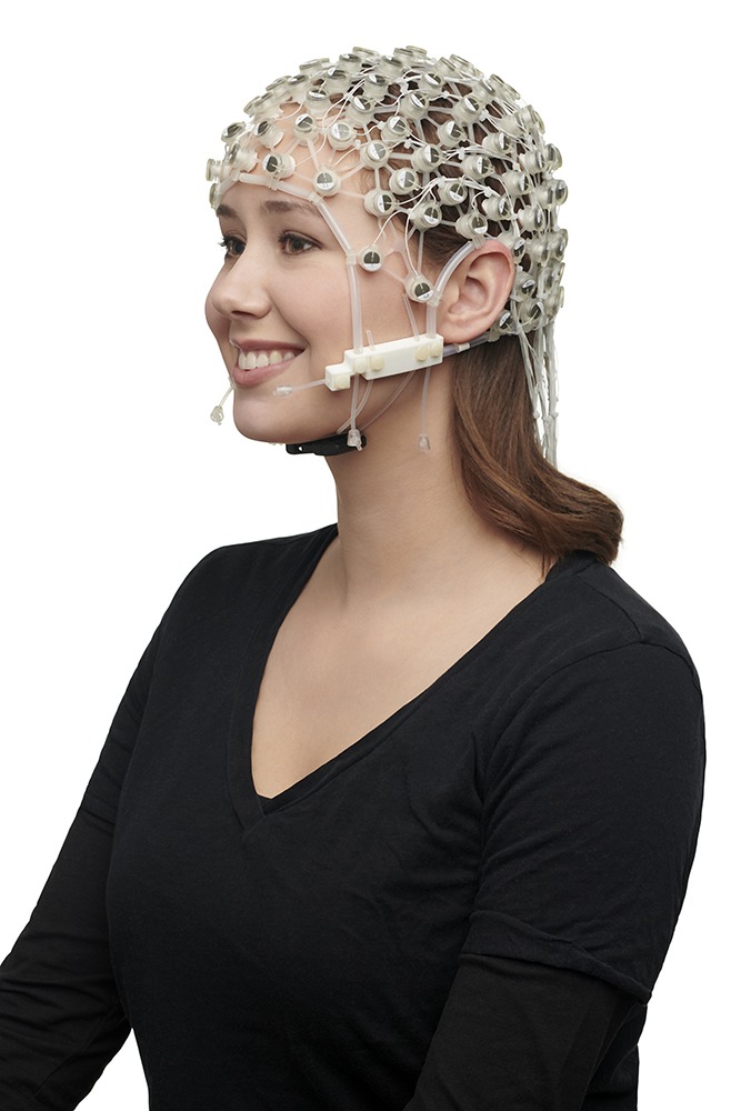
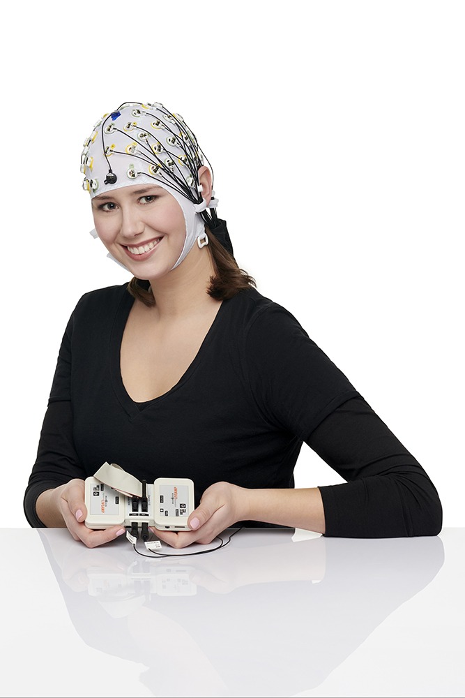
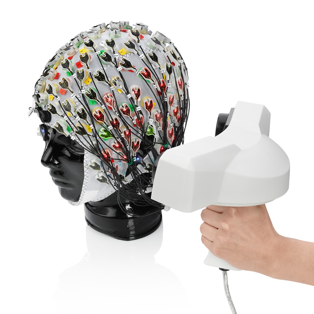
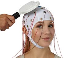
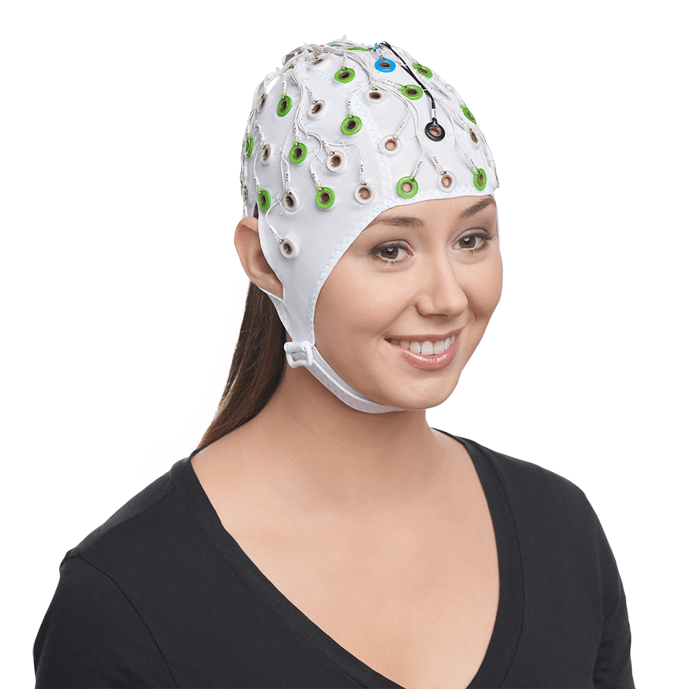
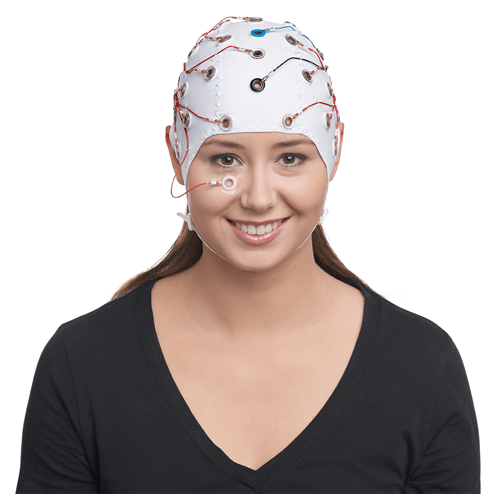
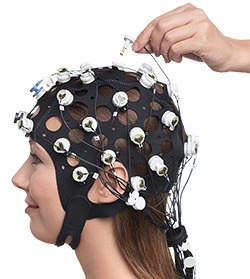
Electroencephalography (EEG) is the measurement of the electrical activity of the brain through specialized electrodes placed on the head. We offer passive, active, gel-based, saline, and dry-electrode technologies, so researchers have flexibility in choosing the right recording electrode for their signals of interest. Often these electrodes are placed on the surface of the scalp and their positions are secured by caps, nets, or headgear. In special cases, electrodes may be placed underneath the scalp or directly on the surface of the brain in a form of electrophysiological monitoring known as electrocorticography (ECoG). Regardless of the approach taken, these electrodes measure the summation of post-synaptic potentials from many brain cells (i.e., neurons) and the resulting waveforms are amplified, digitized, and recorded to better understand brain function under a variety of conditions. We specialize in providing reliable and robust solutions to streamline the measurement and recording of EEG so that researchers can confidently deploy this methodology to address their scientific questions.

EEG Related Products

A black plastic EEG headset (the X.on) floats in the picture. It has seven electrode positions with white sponges for recording EEG data. As a wireless device it can be used in non-traditional recording locations.
The X.on is a mobile EEG platform that can be used with PEER to collect ERP data in places and populations you never would have imagined. It delivers research quality data at a more affordable price point.

X.on + PEER

GrafiCap Dinosaurs design (G113, by Ohmz) is pictured on a mannequin. The cap has 32 green actiCAP snap holders embedded in the fabric and a fun dinosaur pattern can be seen between the holders for fun EEG recordings.
Make EEG recordings fun for your younger participants by using caps with playful printed patterns!

GrafiCap

Here we have a set of B18A snaptrode electrodes fitted into the green actiCAP snap holders of an actiCAP snap cap. The picture is taken from the side of the head showing a part of the mannequin's ear and face. The actiCAP snap logo is visibly printed on the cap fabric.
Pair your existing actiCAP Snap Caps with the new B18A Snaptrode passive electrodes for passive recordings when needed!

Snaptrode (B18A) Bundles

A participant sits wearing an EEG cap while a TMS coil is held to the side of their head. Behind them are two computer screens showing realtime EEG data as recorded and processed by the actiCHamp Plus, TurboLink, and bossdevice RESEARCH that sit on the table.
An accessible platform for brain-state-dependent brain stimulation.

bossdevice RESEARCH with bossapp RESEARCH

Accessing actiCHamp (Plus) data within less than 1.5 ms

TurboLink

The Quick-32r is a complete rethinking of the capabilities of a dry EEG headset. With industry-leading signal quality, built-in noise rejection, long-term comfort, enhanced usability, and a unique built-in LED impedance indicators for real-time contact quality.

Quick-32r

Whether you are aiming for unlocking mechanisms of brain connectivity and function, or for enhanced diagnostic use on epileptiform data, BESA Research 7.1 will help you to achieve your targets reliably and with more pathways than ever before.

BESA Research 7.1

SIGGI-2 is a lab tool with parameters in the micro-volt range.

SIGGI-2

This cap is equipped with actiCAP SNAP holders for the actiCAP slim electrodes, for use with the actiCAP / actiCHamp system.

Cap with actiCAP SNAP holders (for actiCAP Slim electrodes)

The BrainCap for Sleep is a complete cap with built-in electrodes for EEG, EOG and EMG recordings, optimized for sleep recording situations.

BrainCap-SLEEP

A central control unit from a Codamotion system with combined power and data connections for up to eight CODA sensor units.

CodaHub

MREyes is a novel solution that allows operators to see, and to record, a clear view of the subject’s eyes. This can be used in applications as simple as verifying if the eyes are open or closed, or in watching alertness or changes in gaze.

MREyes

The Transcranial Focal Stimulator is designed to be compatible with our unique concentric ring electrodes.

Transcranial Focal Stimulator

The outer ring and the center disc are used for delivering focal stimulation.

Concentric Ring Electrode (CRE) for Stimulation

The t-Interface serves to configure the TCRE electrode elements, route, and pre-amplify the signals.

t-Interface 20 | v2.0

The TCRE performs the Laplacian automatically and takes bipolar differences of the surface potentials from its three closely spaced electrode elements.

t-Lead: Tripolar Concentric Ring Electrode (TCRE)-based sensor

The Dev Kit includes everything you need to undertake EEG experiments and custom hardware development: an 8-channel amplifier, inputs for CGX dry electrodes and ExG’s, and a headband for mounting dry electrodes.

DEV KIT

Our newest amplifier has 20 channels and is small enough to fit in your hand. Amplifier includes standard DB-25 connector for use with off-the-shelf wet caps.

CAMP Compact Amplifier

Connect to eye trackers and other recorders without the need for software or algorithmic timing compensation.

Wireless StimTrigger

The AIM Physiological Monitor is a compact, sophisticated unit that adds heart rate, temperature, respiration, GSR, PPG/HRV/SpO2 and more to any EEG system.

AIM Physiological Monitor

A quick-install wet-cap based system with active electrodes in a standard 10-10 (64-Channel) or 10-5 (128-Channel) configuration, all with a detachable amplifier for the freedom to conduct even the most demanding experiments.

MOBILE-128

Front view of the Quick-20r EEG headset with labeled electrode positions
The Quick-20r v2 is a complete rethinking of the capabilities of a dry EEG headset. With industry-leading signal quality, active electrodes for unparalleled noise rejection, long-term comfort, enhanced usability, and a unique on-board impedance checking system for instant confirmation.

QUICK-20r v2

The Wireless Trigger provides a fully mobile solution for recording event markers in EEG data.

Wireless Trigger

The USB 2 Adapter (also known as BUA) serves as a USB interface to connect any BrainAmp amplifier with the recording computer.

USB 2 Adapter (BUA)

The TriggerBox helps to handle and merge triggers arriving from different sources, which is quite often a requirement in complex laboratory setups.

TriggerBox / TriggerBox Extension

StimTrak combines creativity and precision, making it a very versatile tool which is destined to become a must-have for any kind of laboratory.

StimTrak

Black photo sensor cable from Brain Products, featuring a circular sensor head with transparent window and a long coiled cord ending in a connector
Non MR compatible sensors for peripheral signal recordings

Sensors non MR compatible

The Brain Products Respiration Belt is used to measure the thoracic or abdominal respiratory movements.

Respiration Belt

The new BrainVision Video Recorder Kit uses a high-resolution network camera, integrated with BrainVision Recorder to provide you with excellent synchronization of your video and EEG recordings.

Recorder add-on: BrainVision Video Recorder Kit

The R-Net is based on saltwater sponges and passive Ag/AgCl electrodes that are held in place with a durable and flexible silicone structure.

R-Net: The EEG Net from Brain Products

Rechargeable battery to be used in the lab and the MR environment

PowerPack

Black photo sensor cable from Brain Products, featuring a circular sensor head with transparent window and a long coiled cord ending in a connector
The Photo Sensor is a valuable tool to check the performance of a monitor or any experiment control software. This becomes essential if visual stimuli are planned to be presented on a tablet or cell phone display.

Photo Sensor

Black EEG cap with integrated electrodes connected via cable to a white Brain Vision LiveAmp amplifier unit, displayed on a neutral mannequin head
The LiveCap is a fully customizable cap, especially designed for use with LiveAmp amplifiers to provide a highly comfortable mobile EEG recording solution.

LiveCap

Top-down view of the Brain Vision LiveAmp Sensor and Trigger Extension (STE) module. The device features eight labeled AUX input ports, Trigger In and Out ports, DC Out, and Power In connections, all housed in a compact white rectangular unit
Combining the LiveAmp with the LiveAmp Sensor and Trigger Extension (STE) makes it possible to add additional physiological signals to your mobile EEG research.

LiveAmp Sensor & Trigger Extension (STE)

Close-up view of the Brain Vision LiveAmp device, a compact white portable EEG/ExG amplifier with labeled ports for Trigger and AUX/PWR. The unit features LED indicators and a power button on the top panel
The LiveAmp is a wearable, 24-bit amplifier – available with 8, 16, 32 and also with 64 channels. As it is wireless and allows you to store your recorded data internally (i.e. on an exchangeable memory card), there are no mobility limitations.

LiveAmp 8 / 16 / 32

A woman wearing a white EEG cap with multiple electrodes (actiCAP snap) smiles while seated at a table, holding the Brain Vision LiveAmp mobile amplifier system with open input ports visible
With the LiveAmp 64, you can record high density and high-quality EEG without mobility limitations.

LiveAmp 64

Close-up of the LabSim device, an 8-bit trigger signal generator used for simulating physiological signals in EEG/ERP research systems. The unit features ports labeled AUX DC, TRIG, AUX, EEG, and a power switch, along with a warning indicating it should not be connected to human subjects
LabSim is a small and handy signal generator, able to generate signals required to test the input ports of different amplifiers.

LabSim

Components of a skin conductance (GSR) measurement setup arranged side by side, including adhesive electrode stickers, paired electrodes, a GSR amplifier module, a connecting cable, and a bottle of skin conductance electrode paste
Add this valuable accessory to your already existing BrainAmp ExG MR amplifier and record GSR of laboratory-standard quality even during functional MRI!

GSR MR Sensor

Side view of P CapTrak EEG cap on mannequin head with multicolor electrode layout, shown with TMS coil positioned for compatibility demonstration
CapTrak is a light, hand-held scanner equipped with two integrated cameras and an intuitive, easy-to-use software for fast electrode localization.

CapTrak

P_BrainCap_TMS worn by a participant with a TMS coil positioned over the scalp for simultaneous EEG and TMS recording
This solution allows the TMS coil to be placed very close to the scalp and elicits the evoked response with low magnitude stimulus.

BrainCap TMS

Partial side view of the P BrainCap MR worn by a person
Superior solution for combining EEG & fMRI

BrainCap MR

Frontal view of the P BrainCap MEG on a model with visible multirodes
The BrainCap MEG is a complete configured cap with built-in electrodes for EEG Recordings with simultaneous MEG.

BrainCap MEG

Frontal view of the P BrainCap worn by a person
The BrainCap makes the preparation time very fast and assures high subject comfort as well as high-quality data recordings.

BrainCap

Rendered view of BrainAmp ExG MR PowerPack with ExG AUX
The BrainAmp ExG MR bridges this gap and makes the recording of bipolar and polygraphic signals in the MRI environment a simple procedure.

BrainAmp ExG MR

Rendered view of P BAMR BUA PP
The first BrainAmp MR amplifier for EEG/fMRI simultaneous acquisition was manufactured by Brain Products more than a decade ago and represented a real technological breakthrough.

BrainAmp MR

Rendered view of the P BAST PowerPack
The BrainAmp amplifier is a compact solution for neurophysiology research that can be combined with other units within the same product family to cover a vast range of possible application areas.

BrainAmp Standard

Rendered view of the P BAMRp BUA PP
The standard in combined EEG & fMRI recordings

BrainAmp MR plus

Rendered view of the P BA ExG PP AUX
The BrainAmp ExG amplifier is a bipolar amplifier which can be used to record EMG, ECG, EOG and polygraphic channels such as GSR (Galvanic Skin Response), acceleration, temperature and blood pulse with an extremely compact design.

BrainAmp ExG

Rendered view of the P BADC PowerPack
The BrainAmp DC includes all the outstanding features of the BrainAmp amplifier with the addition of the DC recording mode as well as with multiple hardware filtering options.

BrainAmp DC

P Application Suite trial kit by Brain Products
In order to make it easier and more convenient to evaluate our software solutions for EEG/EP recordings and analysis we are introducing a new trial concept, the so-called “Application Suite Trial Package”.

Application Suite Trial Package

P Analyzer charts
The analysis software for EEG/ERP research

Analyzer 2

Rendered view of P actiCHamp Plus
The actiCHamp Plus can be combined with all our active as well as passive electrode systems – incl. the R-Net, and is powered by a sturdy lithium-ion battery, the PowerUnit.

actiCHamp Plus

Rendered view of P actiCAP Xpress Twist
actiCAP Xpress Twist: dry, fast & comfortable

actiCAP Xpress Twist

Rendered view of P actiCAP snap for LiveAmp
The actiCAP is the go-to solution for reliable and flexible high-quality EEG recordings.

actiCAP slim / actiCAP snap

Rendered view of the P Acceleration 3DMR connectors
MR usable sensor to capture movements in 3 dimensions

Acceleration Sensor MR (3D)

Frequently Asked Questions

Many researchers have asked us this question. What we have found is that impedance matching pastes and gels are not direct shorts like wires. We have measured the conductance of impedance matching pastes and gels and found that they are typically about the conductance of the scalp. Therefore, a millimeter separation of the tripolar electrode elements provides enough resistance in the paste that currents flowing through it cause a potential difference (Ohm’s law: V=IR). These potentials cannot be measured with conventional EEG amplifiers, so we designed our own preamplifier, the t-Interface 20, to go between the t-Leads and the conventional EEG amplifier.

The gain for the tEEG (tripolar EEG) signal from the t-Interface 20 is 187.5. There is no gain for the eEEG (emulated conventional EEG) signal.

No. The t-Interface 20 has been designed specifically to be compatible with your current amplifier and software. The outputs of the t-Interface 20 are standard touch-proof connectors that mate with most common amplifiers on the market today.

We only use the ground signal in our t-Interface 20. If your amplifier uses a reference signal, then it is necessary to connect a reference disc electrode to the subject and the amplifier. If your amplifier does not use a reference signal, then you only need to apply a disc electrode to the subject and connect to the ground input on your amplifier. In either case, you also need to connect the t-Interface 20 ground (Green wire) to the ground input of your amplifier. Prepare and place the disc electrodes as you normally would, and connect them to your amplifier as you normally would. If you do not have two connected isolated ground ports on your amplifier, you will need to use a jumper to connect both the ground conventional disc electrode and the Green wire of the t-Interface 20 output cable to ground on your amplifier. We do not include conventional disc electrodes with our system.

Yes. We typically use Ten20 paste.

You can restock consumables and purchases accessories for your system using the Brain Vision webshop:

Brain Vision LLC (US): https://shop.brainvision.com/

Brain Vision Solutions Inc (Canada) http://shop.bv-solutions.com

Setting up t-Leads on the head for a recording using conductive paste takes no more time than setting up conventional disc electrodes on the head for a recording using paste.

There are no conventional EEG amplifiers, that we are aware of, that have adequate specifications to acquire signals from the t-Leads.

Further, the differencing of the outer ring and central disc, and middle ring and central disc of the t-Lead, along with the Laplacian algorithm first reported in Besio et al. 2006, is performed within the t-Interface 20 circuitry. Therefore, t-Leads will not work with a standard amplifier and you will not get the benefits of the automatic Laplacian provided by the tripolar signal.

The t-Interface 20 is where the differencing between the rings of the t-Leads occurs. Since there is only one ring/sensor on conventional disc electrodes, there is no need to plug disc electrodes in to the preamplifier. Further, our unique TCRE design and t-Interface 20 provides an equivalent conventional disc electrode output for each t-Lead. The t-Interface 20 provides both the high-fidelity tEEG (tripolar EEG) and eEEG (emulated conventional EEG) simultaneously from the each t-Lead.

Additional Application Fields and Solutions

Website Search

Schedule a call with our Consultants​

Please select your area to schedule an introductory call with one of our Scientific Consultants.

script here