The technique of combined EEG & fMRI recordings has been evolving constantly over the last years. The electrode cap and electrodes, which are connected directly to the subject, are the items in our MRI product portfolio that more than the others require constant modifications, in order to guarantee always the highest safety and comfort of the test subject as well as to ensure an outstanding data quality both from the EEG and MRI point of view.
Guaranteed Safety
- All the electrodes in the BrainCap MR are fitted with serial current-limiting resistors.
- Safety resistors are also placed inside the cap connector, acting like an additional RF-filter.
- Electrode cables are routed on the outside of the cap and secured to it so that loops are not formed and cable movement is avoided.
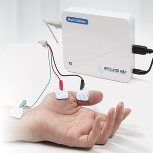
- The drop-down electrodes (e.g. ECG, EOG, EMG) are additionally sheathed in plastic to avoid direct contact with the skin of the test subject.
- ECG electrodes do contain higher resistors than normal electrodes to compensate for the technical characteristics of longer wires.
- Wire length from electrode to the amplifier input is fixed to the minimal required length.
- Wire outlets for the cable tree at central positions avoid loops due to cable routing.
Guaranteed Comfort:
- Flat electrode holders are used to guarantee the comfort of the cap, especially when the head of the test subject in supine position is resting on the electrodes.
- Spare electrode holders are added to caps with less than 64 channels to compensate for gaps between the electrodes. This increases the number of contact points between the test subjects’ head and the MRI scanner head rest to further decrease discomfort for the test subject.
- Soft cap fabric increases comfort and widens the fit to accommodate different head sizes.
Ease of Use
- Serial number sticker for easy tracking, assembly and layout questions.
- Colored electrode holders for easy access/clarification of assembly affiliation (e.g. 10-20, 10-10).
- Name labels on every electrode for easy recognition.
What happens to old electrode caps that do not meet the current safety standards?
If you are sure that your scanner equipment has not changed and that scanner sequences and related procedures are exactly the same as when you first received your equipment, there is no urgent need to change your caps. Otherwise, after a couple of years of use it may be a good idea to adapt your equipment to the latest safety standards. Please verify if your scanner has been upgraded or sequences have been added by the manufacturer.