Trademarked Brain Vision blue icon representing Peripheral Physiology Activity

Peripheral Physiology Activity

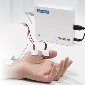
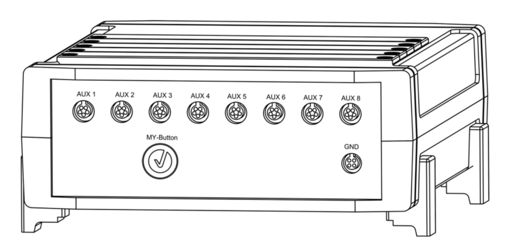
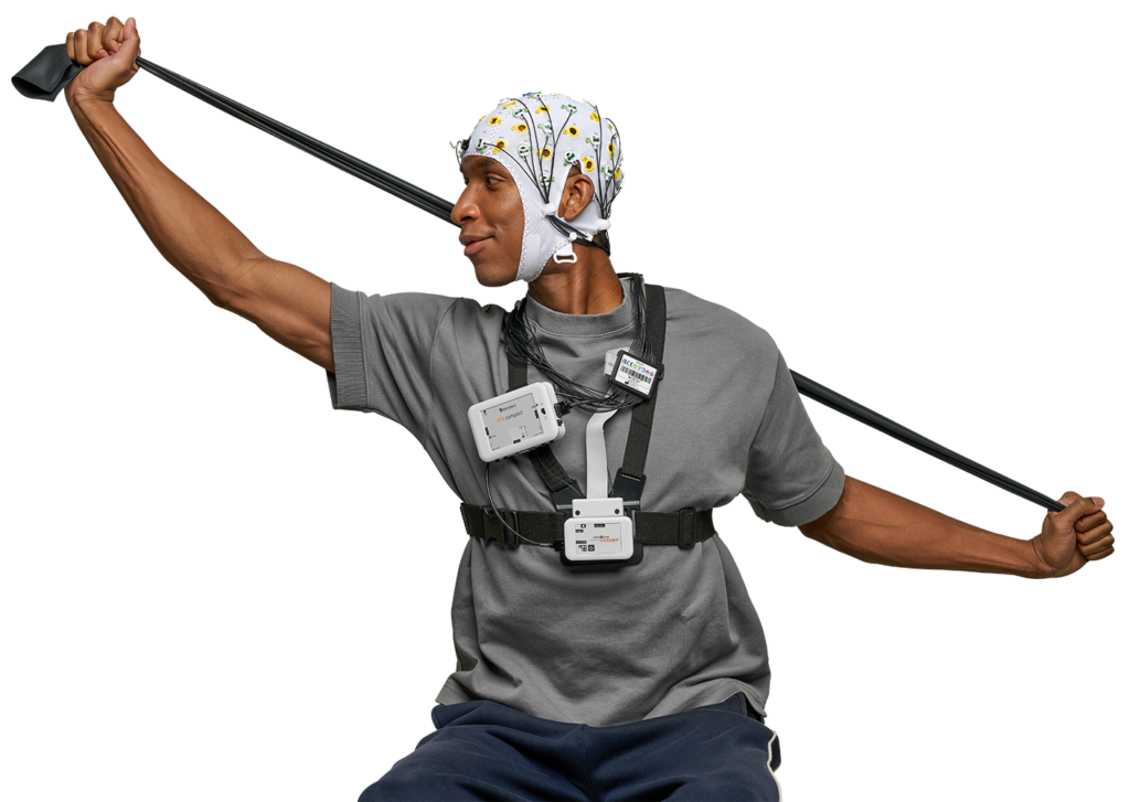
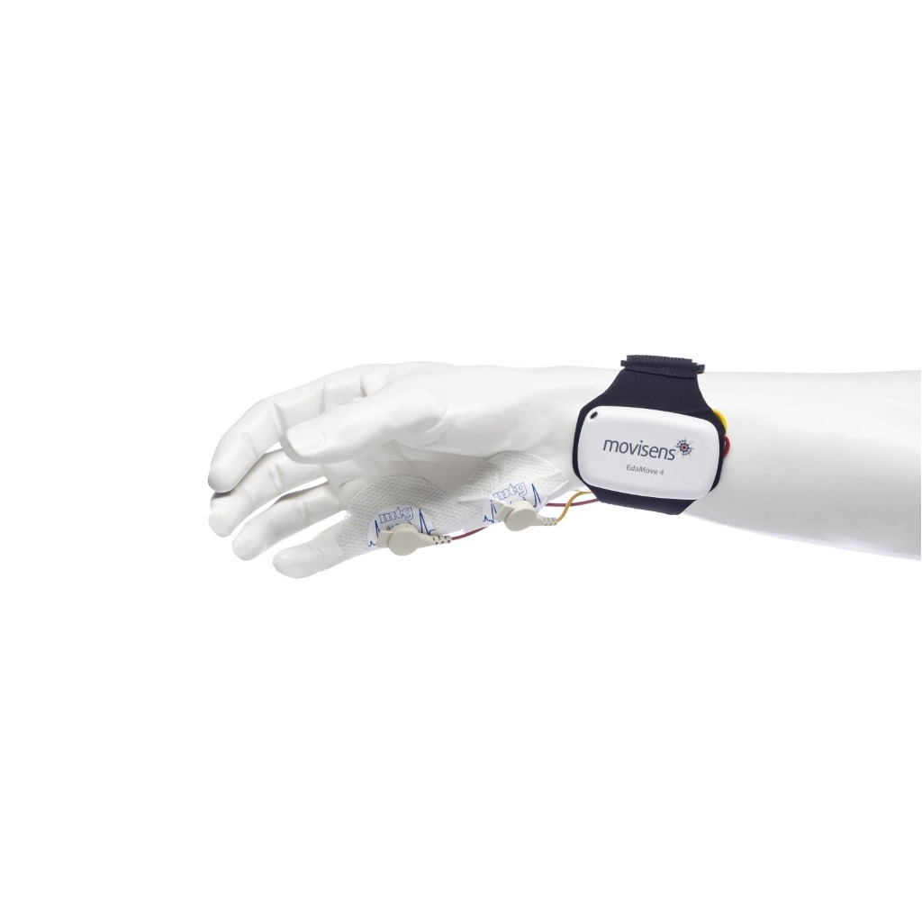
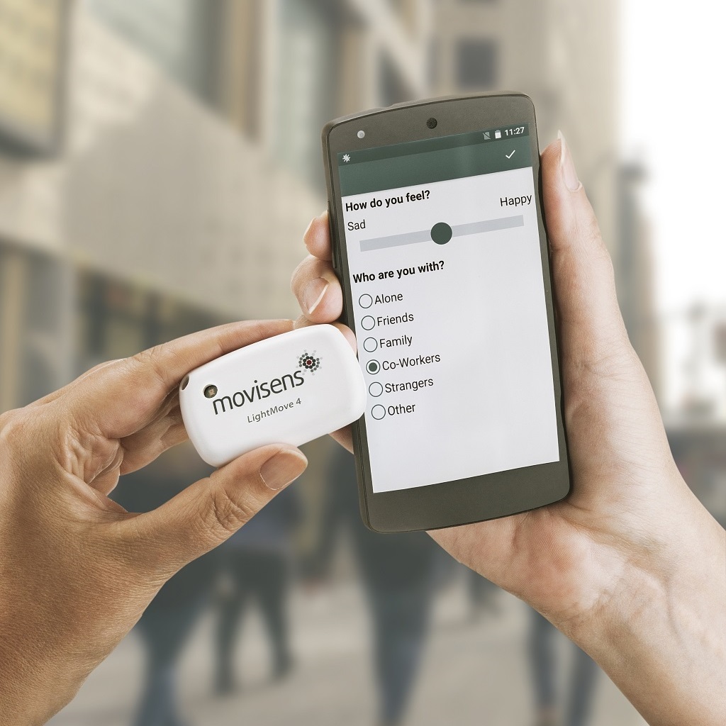
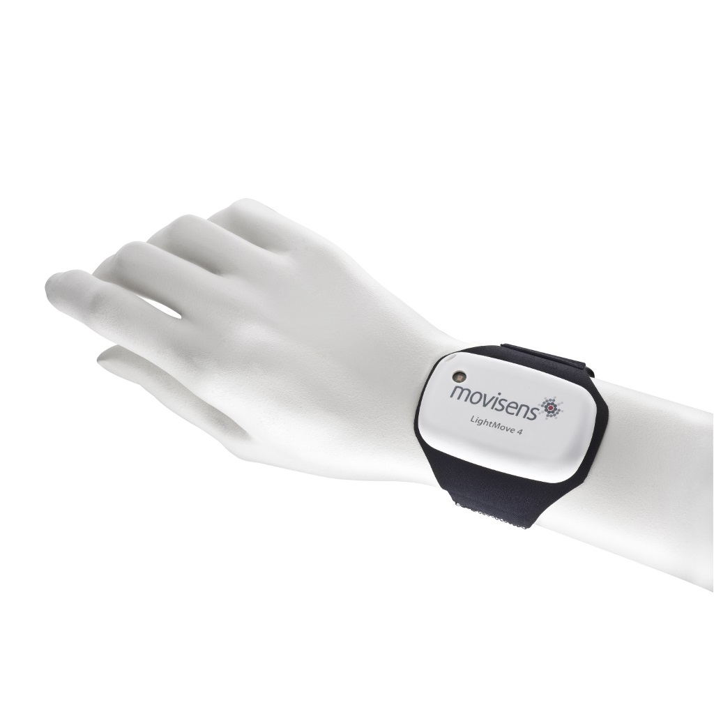
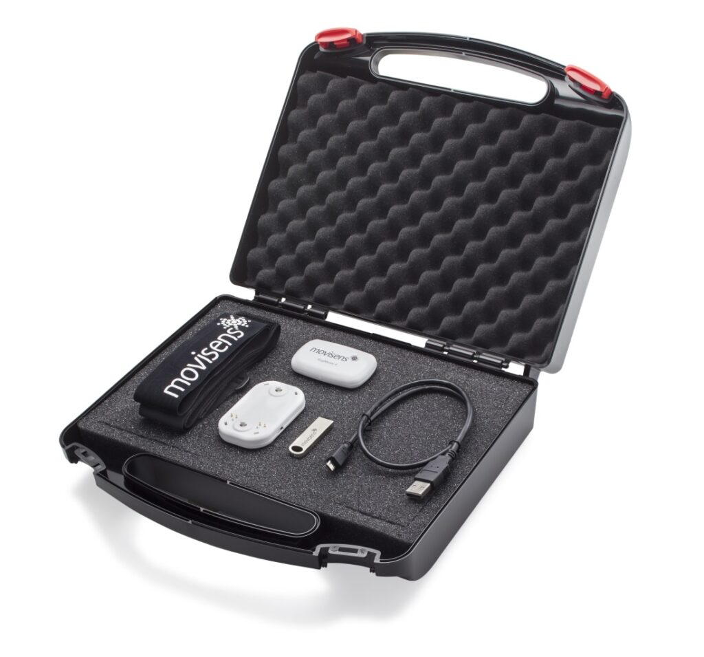
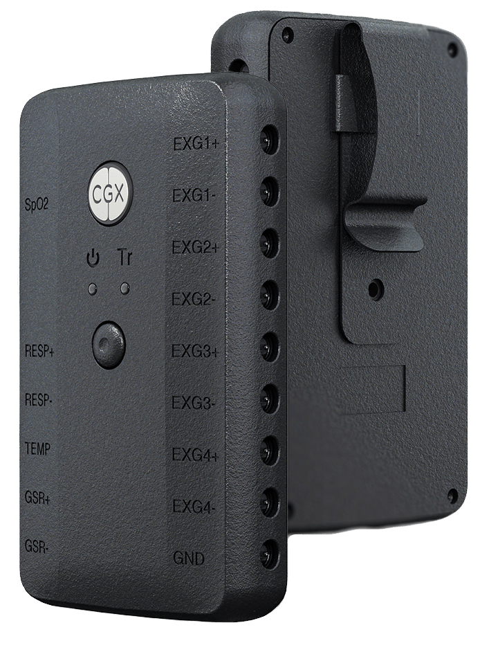
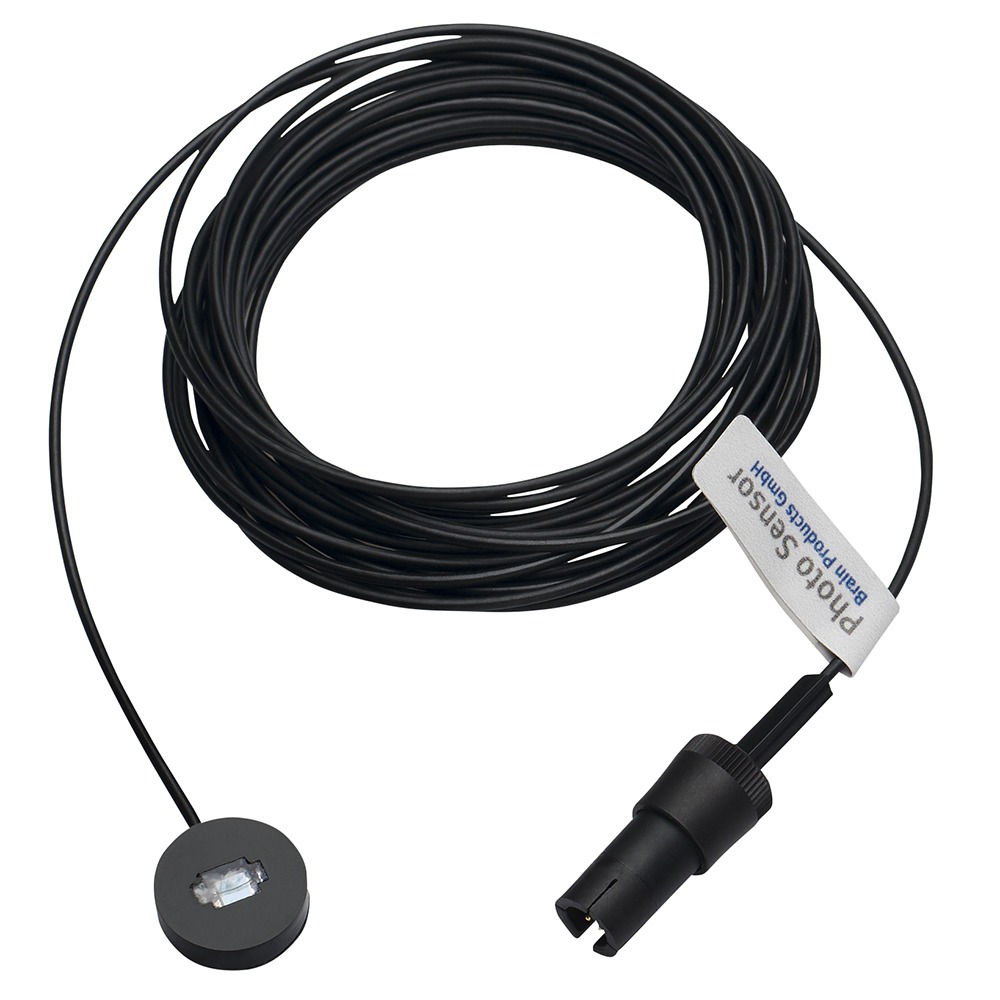
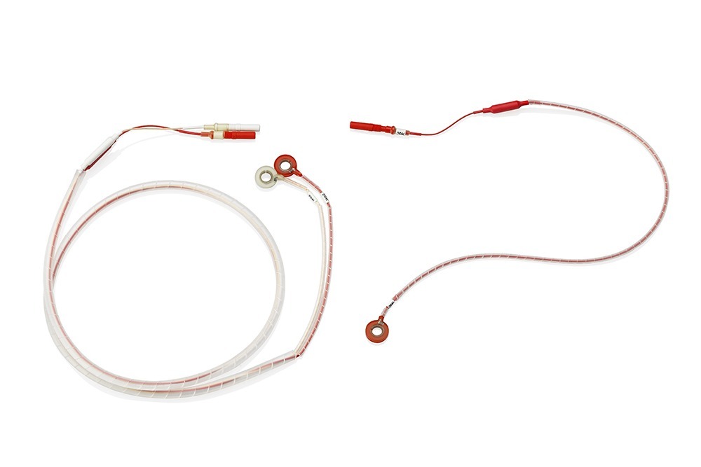
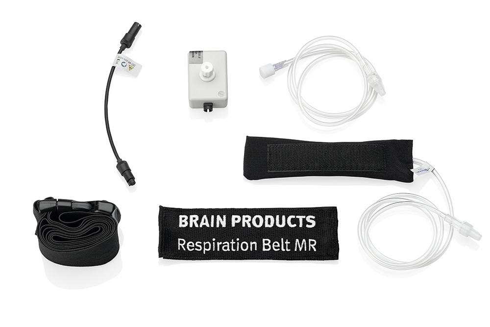
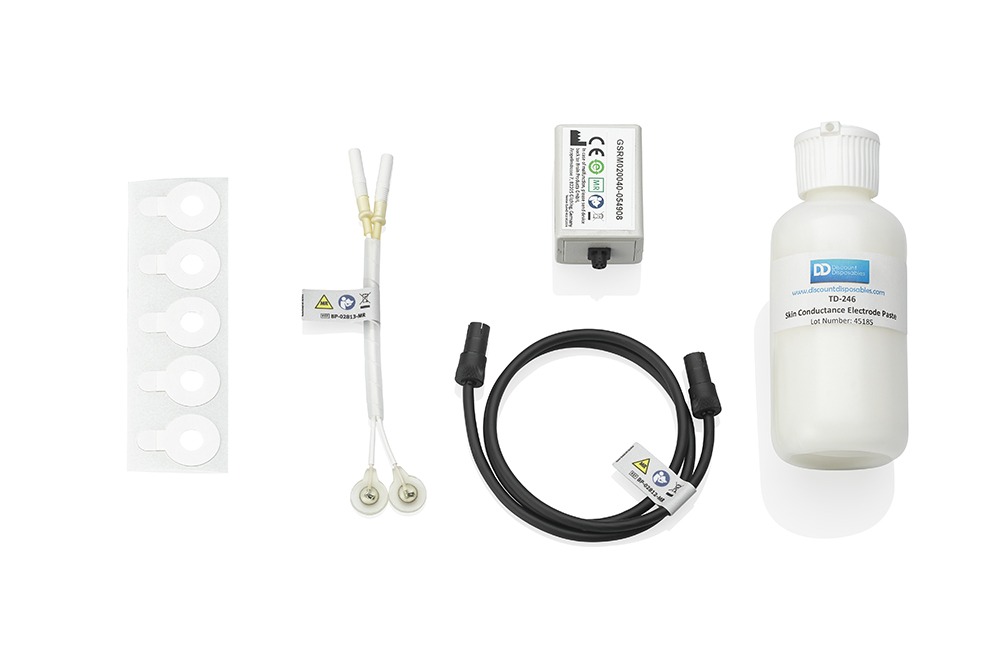
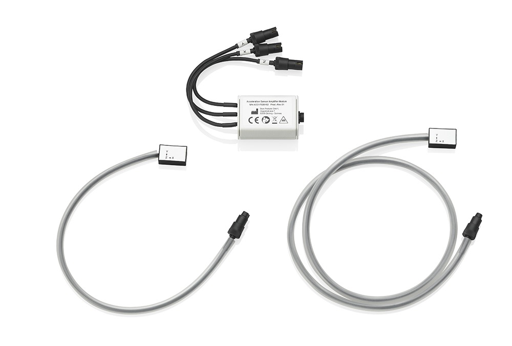
Peripheral physiology can be used to infer activation of the autonomic nervous system in response to various cognitive processes. Peripheral signals (e.g., heart rate [ECG], muscle activation [EMG], eye movements [EOG], skin conductance [GSR], respiration, acceleration, pulse oximetry, etc.) are measured by specialized electrodes and/or sensors that are placed along the body. These signals can be informative on their own, or they may be combined with EEG to inform the interpretation of the resulting waveforms. The requirements for recording clean peripheral signals often differ from those for recording clean EEG signals. For example, peripheral signals tend to be larger (e.g., on the order of mV) compared with EEG signals (e.g., on the order of µV), so there are important technical considerations to keep in mind when selecting an appropriate system for a specific research program. We offer several solutions (including amplifiers, accessories, and open-ended auxiliary cables for researchers to incorporate their own sensors) for recording peripheral physiology that can be used independently or in conjunction with EEG to answer a wide array of questions about psychophysiology. Being able to record peripheral physiology and EEG in the same data file, synchronized to the onset of specific stimuli, streamlines data pre-processing and interpretation of the results.

Peripheral Physiology Activity Related Products

The Brain Ultimate MEP Module for M100 TMS System. In this picture a MEP module is a while box with electrode leads coming out of it and attaching across the thumb muscles of an out stretched hand.
Directly measure EMG signals with your Brain Ultimate M-100 TMS system to quickly and easily identify the motor evoke potential (MEP) threshold when calibrating the TMS pulse magnitude for individual participants.

Brain Ultimate Dual-channel EMG MEP Module

Force Sensor is depicted between a finger tip and a button on a computer keyboard to allow for precise measurement of when force was applied by the finger.
Measure relative changes in force (Newtons) alongside your EEG or other peripheral physiology measures.

Force Sensor

An actiCHamp Plus AUX is drawn by hand showing its front eight (8) AUX ports ready but no EEG channel modules installed.
An expandable platform for peripheral physiology today and up to 160Ch of EEG + 8Ch of AUX tomorrow!

actiCHamp Plus AUX

A male participant is depictred doing stretching with an exercise band while wearing an actiCAP snap EEG cap and a chest harness across his front that holds a LiveAmp, actiCAP slim electrode bundle, and a LiveAmp Sensor & Trigger Extension Compact (STEc).
Expand the capabilities of your LiveAmp to record 6x AUX, wireless triggers, and pulse oximetry with the STEc.

LiveAmp Sensor & Trigger Extension Compact

As part of the new sensor generation, the EdaMove 4 takes the proven quality of the EdaMove 3 and incorporates the most sought after improvements from extensive discussions with researchers in the field.

EdaMove 4 – EDA and Activity Sensor

movisens is proud to be the first company to provide a sensor trigger for Experience Sampling and Ecological Momentary Assessment studies

SensorTrigger – Interactive Assessment

The LightMove 4 opens new possibilities for researchers who understand the interplay between light and physical activity, allowing their research to expand and contribute to this growing body of knowledge.

LightMove 4 – Light and Activity Sensor

The EcgMove 4 strengthens it’s position as the go to device for researchers who care about high quality ECG and Activity data.

EcgMove 4 – ECG and Activity Sensor

The AIM Physiological Monitor is a compact, sophisticated unit that adds heart rate, temperature, respiration, GSR, PPG/HRV/SpO2 and more to any experiment.

AIM Physiological Monitor

Black photo sensor cable from Brain Products, featuring a circular sensor head with transparent window and a long coiled cord ending in a connector
Non MR compatible sensors for peripheral signal recordings

Sensors non MR compatible

To meet the demands of our customers we are continuously working on developing sensors with the capability to measure peripheral physiological signals in addition to EEG in the MR scanner.

Sensors for MR

MR usable sensor for parallel respiratory measurements

Respiration Belt MR

Components of a skin conductance (GSR) measurement setup arranged side by side, including adhesive electrode stickers, paired electrodes, a GSR amplifier module, a connecting cable, and a bottle of skin conductance electrode paste
Add this valuable accessory to your already existing BrainAmp ExG MR amplifier and record GSR of laboratory-standard quality even during functional MRI!

GSR MR Sensor

Rendered view of the P BIP2AUX adapter with Multitrodes
The BIP2AUX Adapter is a tiny, analogue differential DC amplifier that amplifies the incoming signal in order to make it measurable and relays it through the auxiliary input ports of the V-Amp and the actiCHamp.

BIP2AUX Adapter

Rendered view of the P Acceleration 3DMR connectors
MR usable sensor to capture movements in 3 dimensions

Acceleration Sensor MR (3D)

Additional Application Fields and Solutions

Website Search

Schedule a call with our Consultants​

Please select your area to schedule an introductory call with one of our Scientific Consultants.

script here