Respiration Belt MR

MR usable sensor for parallel respiratory measurements

Respiration plays a critical role in the MR environment, where it may not only be a confounding factor, but also a source of related artifacts. It can be linked to movement artifacts, physiological alterations, induced field inhomogeneity, or interference with the experimental paradigm. Therefore respiratory effects cannot be ignored (see e.g. Thomason et al. 2005).

Even if advances in data analysis techniques can provide better results at the cost of greater complexity, these results are considerably improved by parallel dedicated measurements of the sources of the artifacts. An efficient method which exploits parallel measurements for artifact correction uses acquired respiratory signals to create a principal regressor, along with other derived regressors obtained with a higher order analysis of the signal itself. This approach is known as RETROICOR (Glover et al., 2000). It is clear that a higher quality and sensitivity of acquired respiratory data will lead to an improved quality of all the regressors and finally to a higher quality of artifact correction and final denoised data, independent of the strategy adopted to correct for respiratory artifacts.

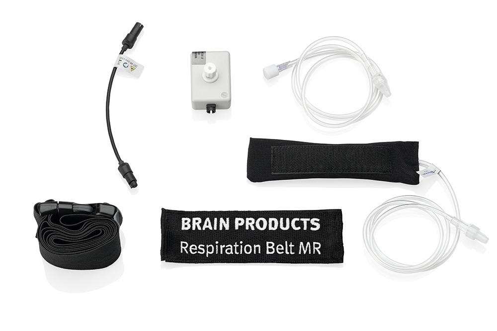
With the aim of obtaining the best data quality and the optimal method of artifact correction we have developed the Respiration Belt MR, a sensor for the acquisition of respiratory signals within MR environments.

Related Products

The Brain Ultimate MEP Module for M100 TMS System. In this picture a MEP module is a while box with electrode leads coming out of it and attaching across the thumb muscles of an out stretched hand.
Directly measure EMG signals with your Brain Ultimate M-100 TMS system to quickly and easily identify the motor evoke potential (MEP) threshold when calibrating the TMS pulse magnitude for individual participants.

Brain Ultimate Dual-channel EMG MEP Module

BrainVision Recorder 2 software mode icons are shown in a row numbered 1 through 4 to illustrate the normal workflow for using the software.
Meet the next generation of EEG research recording software!

BrainVision Recorder 2

Force Sensor is depicted between a finger tip and a button on a computer keyboard to allow for precise measurement of when force was applied by the finger.
Measure relative changes in force (Newtons) alongside your EEG or other peripheral physiology measures.

Force Sensor

A blond female model is facing away from us while wearing the Mindtooth Touch System on her head.
Record research-grade EEG out in the wild and apply state of the art neurometrics calculations on live data.

Mindtooth

View Other Application Fields

Website Search

Schedule a call with our Consultants​

Please select your area to schedule an introductory call with one of our Scientific Consultants.

script here