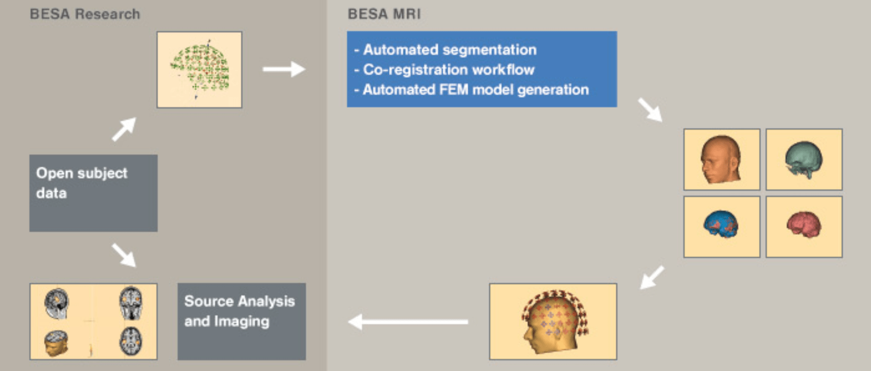
BESA MRI is software to generate individual head models (BEM / FEM) that can be used for EEG and / or MEG source analysis. BESA MRI also allows co-registration of EEG / MEG data with individual MRI data, and visualization of dipole solutions generated in BESA Research in the individual anatomy. BESA MRI is the first software that offers an easy, intuitive interface with an integrated workflow guiding the user step-by-step.
MRI data import is easily performed with DICOM, ANALYZE / NIFTI, VMR readers. The preparation of the MRI data to be automatically processed by BESA MRI requires just a couple of simple work steps typically demanding only a few minutes of user attention. BESA MRI’s automatic processing can be applied to all subjects prepared by the user in one go, therefore conveniently minimizing the time the user has to work on the computer.
BESA MRI’s automatic segmentation includes an automated inhomogeneity correction to correct for scan artifacts, generates a high quality cortex and scalp reconstruction with optional cortex inflation for enhanced visualization, and, in an optional step, can also generate an individual head model (BEM, FEM) with 3 layers (scalp, skull, brain – BEM) or 4 layers (scalp, skull, CSF and brain – FEM).
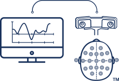
BESA MRI 3.0 runs in combination with BESA Research 7.1 and 7.0. Individual head models (BEM, FEM) generated with BESA MRI 3.0 can be imported by BESA Research 7.x for EEG and / or MEG source analysis. EEG / MEG data can be co-registered with individual MRI data using individually digitized electrode positions or head shape points. As a unique feature of BESA MRI 3.0, 10-10 standard electrodes (including inferior electrodes) can also be morphed with individual MRI data. Co-registered electrode coordinates are immediately available in BESA Research. Thus, source images / localizations can be displayed on the individual MRI even if no individual digitization of electrodes has taken place.
The MRI data can be visualized in a user-defined multi-slice view. Discrete solution files generated by BESA Research can be overlaid over the individual anatomy, as well as one of several available brain atlases.
Contact us for a free trial to test how BESA MRI could fit into the data processing pipeline for your next research project!