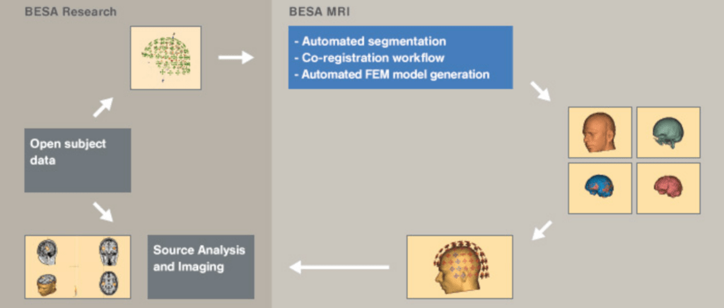
Functional magnetic resonance imaging (fMRI) uses rapidly oscillating magnetic fields to measure changes in blood oxygenation level dependent signaling (BOLD) throughout the brain, which is often considered to be a correlate of neural activation. One strength of fMRI is its high spatial resolution, which permits measurement of activation in small, deep brain structures, whereas EEG is often restricted to the measurement of activation in more superficial areas of the cortex. Because fMRI measures changes in blood oxygenation (i.e., the hemodynamic response), it is a slower signal (on the order of seconds) compared to EEG, which is extremely fast (on the order of milli seconds). The ability to study brain activation with both high spatial and temporal resolution allows researchers to answer more complex questions about neural activation. However, fMRI is traditionally considered to be a hostile environment for EEG. So, from a technical standpoint, it is challenging to combine these two methodologies while maintaining participant safety. We are the market leaders in EEG-fMRI solutions, and we have a dedicated team of fMRI-certified EEG experts to support researchers throughout their studies.