
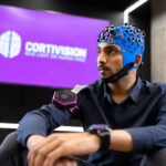
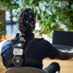
Cortivision PHOTON CAP

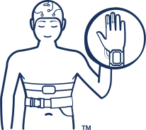
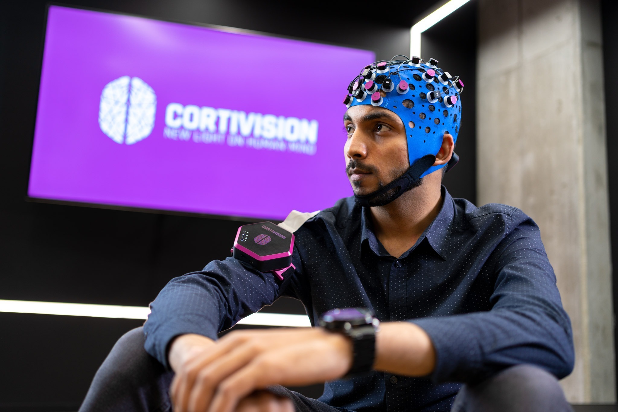
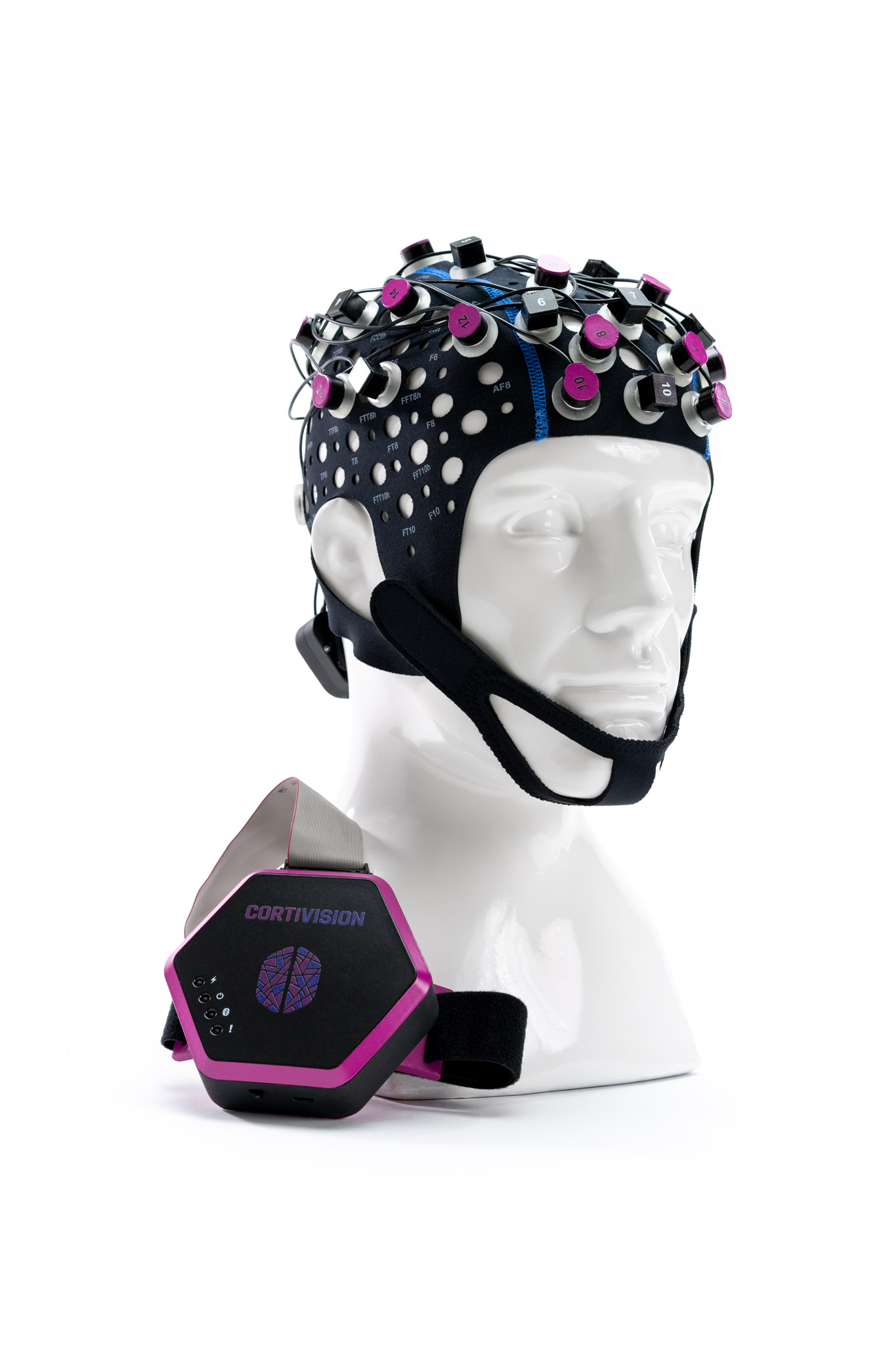
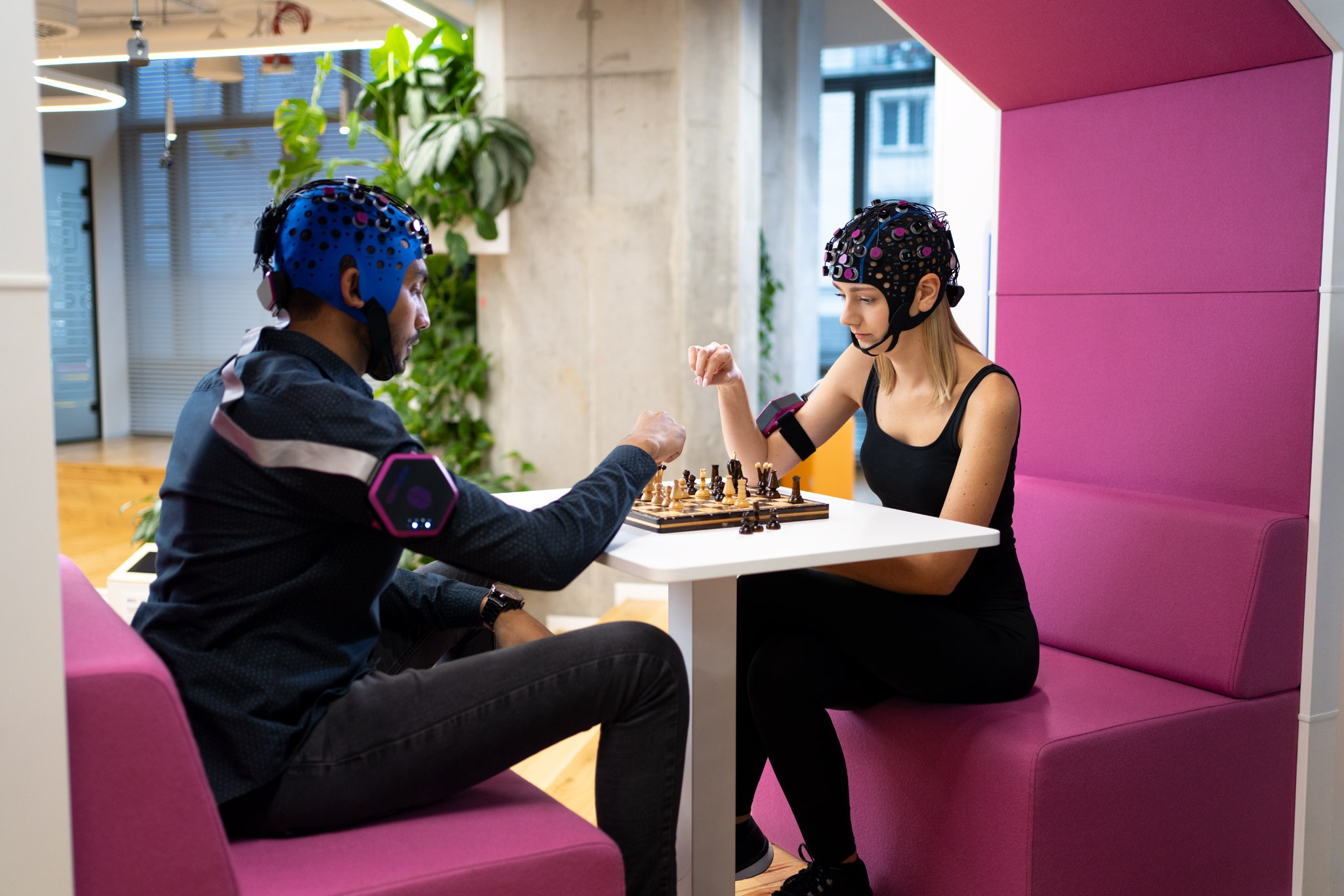
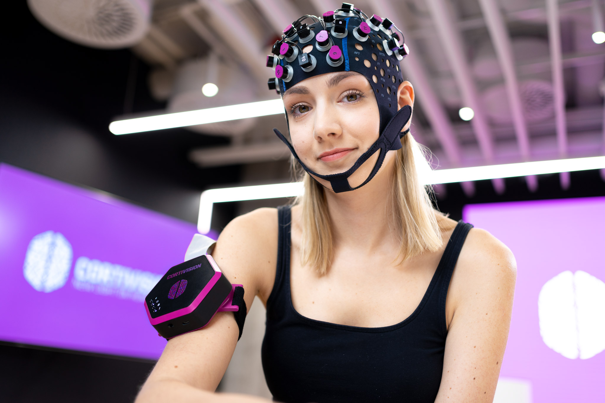
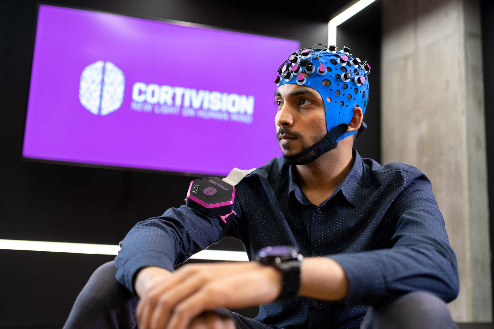
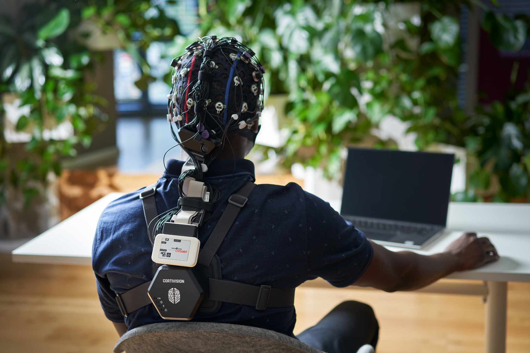
The PHOTON CAP fNIRS system weighs about half a pound (less than 230 grams) and can be worn effortlessly on a subject’s arm or chest while collecting data with its set of 16 sources and 10 detectors. Its ultra-compact design was a key factor in its selection for the Axiom-2 mission to the ISS and in Spring of 2023 it became the first fNIRS device used in space!

With Brain Vision’s experience with fNIRS as well as integrating it with EEG, we are excited to help more researchers apply Cortivision’s solutions in their studies!

Learn more about Cortivision here.
Frequently Asked Questions

Light intensity is heavily attenuated in tissue and falls off exponentially from the illumination point. The maximum achievable probing depth of NIRS is limited by the illumination strength – determined by the thermal damaging threshold – and the detection sensitivity. Imaging depth strongly depends on the tissue type and the application. Typical achievable transmission limits of NIRS are about 12 cm for breast tissue, and 6 cm on the arm or leg. For brain imaging, the probing depth of NIRS is about 3 cm.

Diffuse optical tomography is a low-resolution technique owing to the physics of light propagation in scattering media. Depending on the composition and size of the target tissue, the resolution is on the order of 5-10 mm.

The temporal resolution of an fNIRS device depends on its hardware as well as how its individual channels operate with respect to one another. Through time-multiplexing of the source firing, some systems can achieve anywhere between 3-25Hz depending on the optode montage. For a particular application, scan speed can be traded off against the desired coverage area (field-of-view) or source density (image resolution).

  • Language, Cognition
  • Learning, Memory
  • Sensory, Motor, Visual
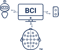
  • Brain-Computer Interface (BCI) Neurofeedback
  • Acute Care, Ischemia
  • Autism
  • Animal Imaging: Rats
  • Animal Imaging: Monkeys
  • Child studies
  • Traumatic Brain Injury (TBI) Studies
  • Multimodal studies NIRS used simultaneously with EEG, TMS, eye-tracking, tDCS, and other modalities.
  • Behavioral studies
  • Motorcontrol & movement related studies
  • Sports performance studies

Related Products

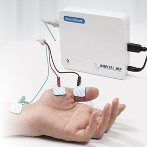
The Brain Ultimate MEP Module for M100 TMS System. In this picture a MEP module is a while box with electrode leads coming out of it and attaching across the thumb muscles of an out stretched hand.
Directly record EMG signals with your Brain Ultimate M-100 TMS system to correctly identify the motor evoke potential (MEP) threshold when calibrating the TMS pulse magnitude with individual participants.

Brain Ultimate Dual-channel EMG MEP Module

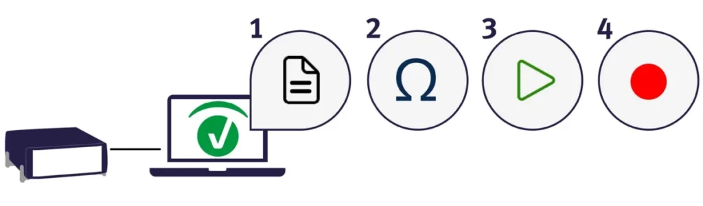
BrainVision Recorder 2 software mode icons are shown in a row numbered 1 through 4 to illustrate the normal workflow for using the software.
Meet the next generation of EEG research recording software!

BrainVision Recorder 2

Force Sensor is depicted between a finger tip and a button on a computer keyboard to allow for precise measurement of when force was applied by the finger.
Measure relative changes in force (Newtons) alongside your EEG or other peripheral physiology measures.

Force Sensor

The LOOP-IT's front panel is depicted with its touchscreen interface, 8x EEG inputs, 2x tES outputs, 4x BNC outputs, and a special port.
The neuroConn LOOP-IT is an all-in-one solution for closed loop EEG-tES, TMS-EEG, and EEG-tFUS.

neuroConn LOOP-IT

View Other Application Fields

Website Search

Schedule a call with our Consultants​

Please select your area to schedule an introductory call with one of our Scientific Consultants.

script here