Trademarked Brain Vision blue icon representing EEG & fNIRS

fNIRS

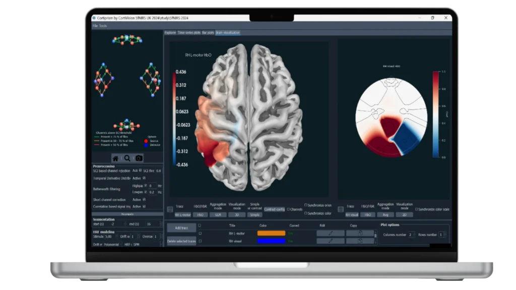
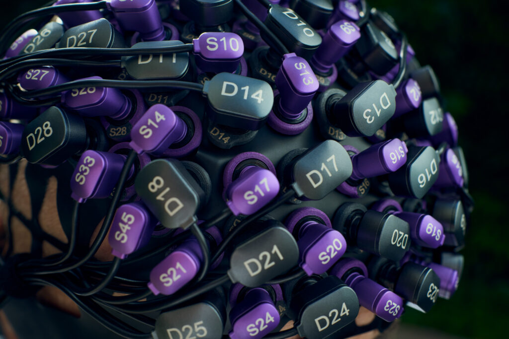
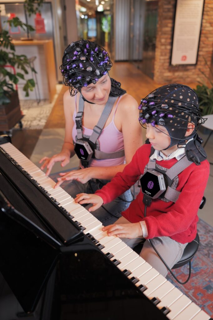
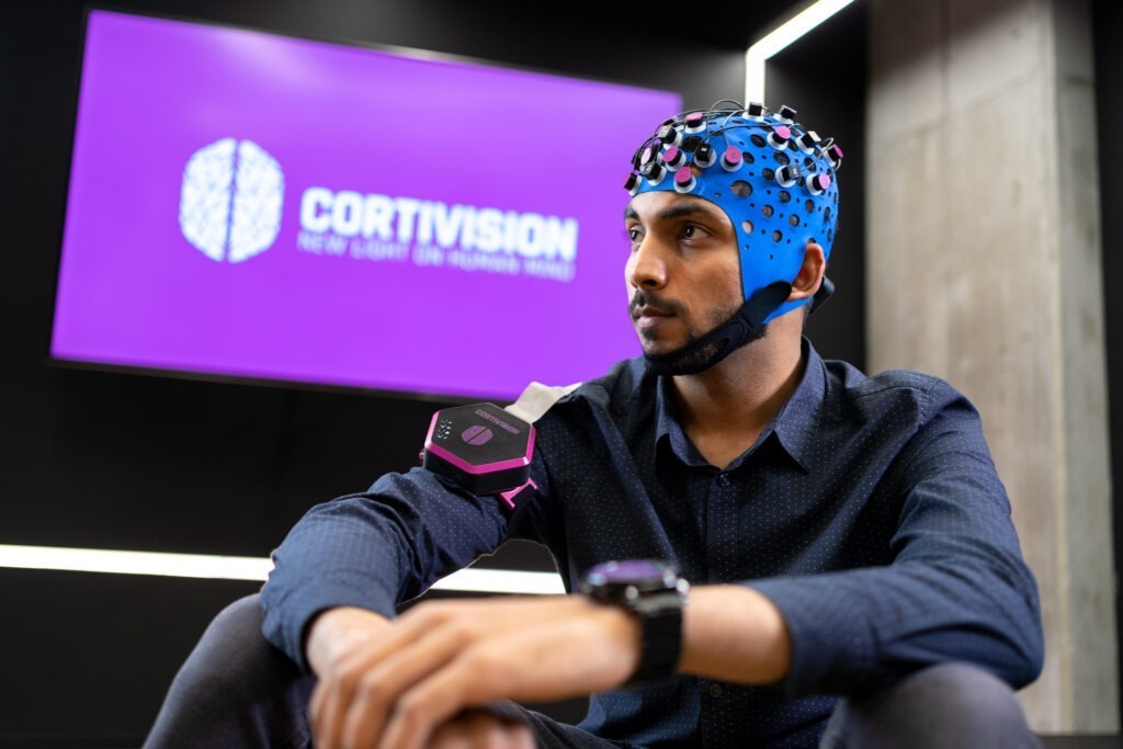
Functional near infrared spectroscopy (fNIRS) uses different wavelengths of near infrared light to measure changes in blood oxygenation and deoxygenation (i.e., the hemodynamic response) across the cortex, which is often considered to be a correlate of brain activation. With fNIRS, specialized optodes are placed on the head, and their positions are secured in a grid-like fashion by caps or other headgear. Near infrared light is then emitted through one set of optodes and directed towards the scalp. Light that is not absorbed by the cortex is measured by surrounding detector optodes. This technique allows researchers to make inferences about brain activation without the need for magnetic fields or currents. Given its portability, fNIRS is versatile and can be used to answer a variety of questions about brain activation under more ecologically valid conditions (i.e., while participants are engaged in real-world activities). Similar to fMRI, it can also be combined with EEG; however, it is limited to cortical brain activation. Compared with EEG, fNIRS is a newer methodology and many labs previously developed homegrown solutions for measuring the hemodynamic response of the brain. We have partnered with leading fNIRS companies to offer cutting-edge, professional solutions that are backed by expert-level support.

fNIRS Related Products

The first fNIRS system used in space, the Cortivision PHOTON has 16 sources and 10 detectors and brings fNIRS recordings to more places outside the lab.

Cortivision PHOTON CAP

A woman and child sit side by side at a piano to play music while both simultaneously wear the wireless and lightweight Cortivision SPECTRUM fNIRS system to record data during a hyperscanning paradigm.
The Cortivision SPECTRUM is a lightweight, wearable, wireless fNIRS platform expandable from 12 sources/8 detectors to up to 48 sources/32 detectors for whole head and DOT imaging.

Cortivision SPECTRUM

Close up image of a Cortivision SPECTRUM DOT cap with high density arrangement of sources and detectors.
A complete hardware and software ecosystem for Diffuse Optical Tomography, from Cortivision.

Cortivision SPECTRUM DOT

CortiPrism provides a robust GUI for analyzing fNIRS data from Cortivision devices in single subject and group level analyses.

Cortivision CortiPrism

BrainVision Recorder is a multifunctional recording software designed to provide our amplifier customers with an extremely versatile and easy-to-use platform for recording setup and execution.

Recorder

Close-up view of the Brain Vision LiveAmp device, a compact white portable EEG/ExG amplifier with labeled ports for Trigger and AUX/PWR. The unit features LED indicators and a power button on the top panel
The LiveAmp is a wearable, 24-bit amplifier – available with 8, 16, 32 and also with 64 channels. As it is wireless and allows you to store your recorded data internally (i.e. on an exchangeable memory card), there are no mobility limitations.

LiveAmp 8 / 16 / 32

A woman wearing a white EEG cap with multiple electrodes (actiCAP snap) smiles while seated at a table, holding the Brain Vision LiveAmp mobile amplifier system with open input ports visible
With the LiveAmp 64, you can record high density and high-quality EEG without mobility limitations.

LiveAmp 64

Rendered view of the P BAMRp BUA PP
The standard in combined EEG & fMRI recordings

BrainAmp MR plus

Frequently Asked Questions

Light intensity is heavily attenuated in tissue and falls off exponentially from the illumination point. The maximum achievable probing depth of NIRS is limited by the illumination strength – determined by the thermal damaging threshold – and the detection sensitivity. Imaging depth strongly depends on the tissue type and the application. Typical achievable transmission limits of NIRS are about 12 cm for breast tissue, and 6 cm on the arm or leg. For brain imaging, the probing depth of NIRS is about 3 cm.

Diffuse optical tomography is a low-resolution technique owing to the physics of light propagation in scattering media. Depending on the composition and size of the target tissue, the resolution is on the order of 5-10 mm.

The temporal resolution of an fNIRS device depends on its hardware as well as how its individual channels operate with respect to one another. Through time-multiplexing of the source firing, some systems can achieve anywhere between 3-25Hz depending on the optode montage. For a particular application, scan speed can be traded off against the desired coverage area (field-of-view) or source density (image resolution).

  • Language, Cognition
  • Learning, Memory
  • Sensory, Motor, Visual
  • Brain-Computer Interface (BCI) Neurofeedback
  • Acute Care, Ischemia
  • Autism
  • Animal Imaging: Rats
  • Animal Imaging: Monkeys
  • Child studies
  • Traumatic Brain Injury (TBI) Studies
  • Multimodal studies NIRS used simultaneously with EEG, TMS, eye-tracking, tDCS, and other modalities.
  • Behavioral studies
  • Motorcontrol & movement related studies
  • Sports performance studies

Additional Application Fields and Solutions

Website Search

Schedule a call with our Consultants​

Please select your area to schedule an introductory call with one of our Scientific Consultants.

script here