BrainAmp MR plus

The standard in combined EEG & fMRI recordings

Since the design of the first BrainAmp MR, the neurophysiology research world moved forward and started to look at new techniques of investigating the brain in action. In light of this, it was obvious that there was a need to develop a more advanced amplifier that could cover a wider range of applications and be as flexible as required by its users.

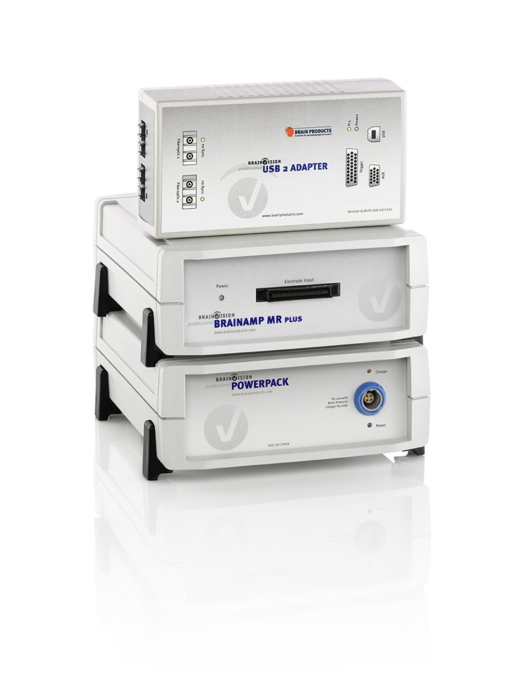
BrainAmp MR plus at a glance

  • BrainAmp MR plus enhances the already outstanding features of the BrainAmp MR.
  • It offers multiple hardware signal resolution options that are easily selectable via the recording software.
  • Depending on the recording needs, with just one “click” it is possible to switch from AC to DC mode acquisition as well as to extend the hardware bandwidth.
  • It’s a shielded amplifier which can be taken directly inside the MRI chamber and placed in the bore right behind the subject’s head.
  • It can be used for simultaneous EEG/fMRI acquisitions as well as for EEG/TMS co-registrations, EEG/ERP studies, and Brain-Computer-Interface applications.
  • The sturdy and compact amplifier is powered by the rechargeable PowerPack battery.
  • Multiple amplifiers can be combined and stacked on top of each other in order to increase the maximum number of available channels to 128 for recordings in the MRI environment and up to 256 channels for laboratory applications.
  • BrainAmp MR plus can be combined with the BrainAmp ExG MR to add the capability to record bipolar and peripheral signals (e.g. EOG, ECG, EMG, GSR – Galvanic Skin Response, etc.) in an extremely compact setup.

There is no need for different amplifier types as the Brain Products’ flagship represents the state of the art solution for all of the aforementioned applications.

Technical Specifications

BrainAmp MR plusTechnical specifications
Amplifier
Number of channels (per unit)32
Channel type / referenceReferential channels/acquisition of a reference using a single electrode (unipolar)
Sampling rate5 kHz per channel
Resolution0.1 μV / 0.5 μV per bit (switchable)
Measuring range±3.28 mV / ±16.384 mV (switchable)
BrainAmp MR plusTechnical specifications
Amplifier
Input impedanceSwitchable: 10 MΩ / 10 GΩ (for DC)
Filter typeFirst-order filter with 6 dB/octave
Lower cutoff frequency (high pass)0 Hz in DC mode or 0.016 Hz/10 s in AC mode; switchable between AC and DC modes
Filter typeFifth-order Butterworth filter with 30 dB/octave
High-cutoff frequency (low-pass)/time constant1000 Hz / 250 Hz (switchable for the resolutions 0.1 μV / 0.5 μV per bit)
Input noise≤ 1 μVpp
Common-mode rejection (CMR)≥ 110 dB
Bit width of the A/D converter (EEG and AUX)16 bit
Amplitude accuracy±2%
DC offset tolerance±300 mV
Integrated impedance measurementPresent; measurement includes ground electrode and reference electrode
Signal transmissionOptically coupled using duplex fiber optic cables
Power SupplyPowerPack (external rechargeable battery)
Power consumption (in operation)Max. 150 mA
Power consumption (in standby)Typically 7 mA
Operating time between chargesTypically 30 hours with one amplifier, 15 hours with two amplifiers
Dimensions (H x W x D)Approx. 68 mm x 160 mm x 187 mm
WeightApprox. 1.1 kg
Unlocking functionPresent
Locking of unused channelsPresent
Suitable for use in MR scanner roomYes

Related Products

The LOOP-IT's front panel is depicted with its touchscreen interface, 8x EEG inputs, 2x tES outputs, 4x BNC outputs, and a special port.
The neuroConn LOOP-IT is an all-in-one solution for closed loop EEG-tES, TMS-EEG, and EEG-tFUS.

neuroConn LOOP-IT

neuroConn DCMC is pictured with its control panel PC, keyboard, and mouse, along with 4x stimulation channels connected to a glass head via rubber electrodes.
An expandalbe multi-channel transcranial electrical stimulation (HD-tES) platform.

neuroConn DC-STIMULATOR MC

neuroConn DC-STIMULATOR PLUS is depicted laying on a table with a pair of rubber/sponge electrodes connected.
A capable and powerful one-channel transcranial electrical stimulation (tES) tool.

neuroConn DC-STIMULATOR PLUS

The BESA Connectivity software interface is a workflow-based GUI that walks you through the connectivity analysis of your choosing. Here you see the results of a Coherence analysis between multiple regional sources (source space, frequency anlaysis).
BESA Connectivity is a stand-alone module that extends BESA Research with streamlined, user-guided workflows for time–frequency and connectivity analysis of EEG and MEG data (in both scalp and source space). With multiple time–frequency and connectivity analysis methods supported, you can run batch analyses across subjects and easily visualize results using a variety of 2D and 3D renderings. The output is also ready for direct import into BESA Statistics.

BESA Connectivity 2.0

View Other Application Fields

Website Search

Schedule a call with our Consultants​

Please select your area to schedule an introductory call with one of our Scientific Consultants.

script here