BrainAmp ExG MR

Bipolar amplifier with unparalleled design for use in the MR environment

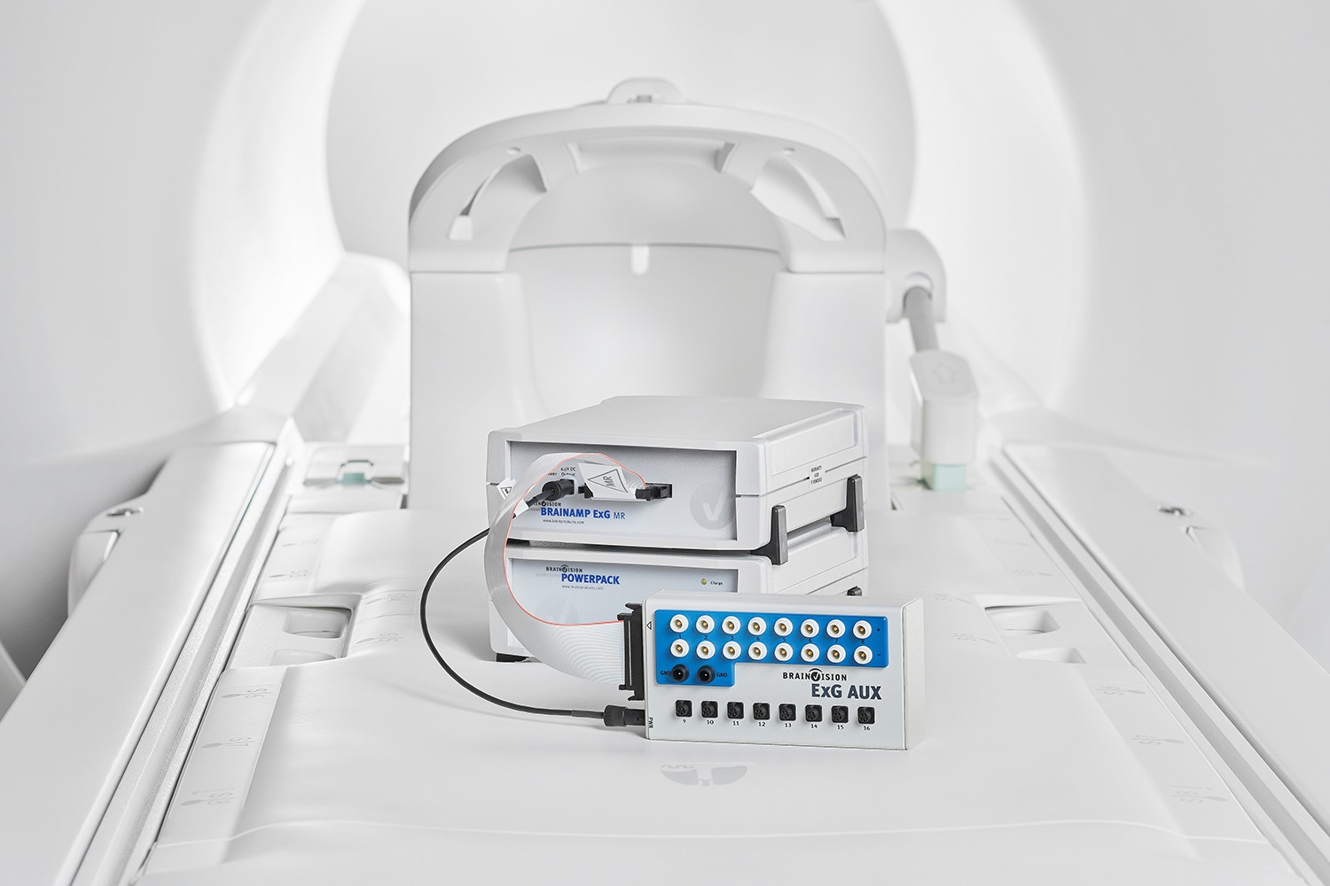
The increased interest in simultaneous EEG/fMRI recordings to investigate brain activity very quickly led to the need to also co-register other types of physiological data such as bipolar and peripheral signals. The BrainAmp ExG MR bridges this gap and makes the recording of bipolar and polygraphic signals in the MRI environment a simple procedure.

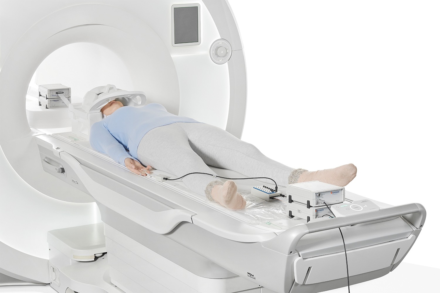
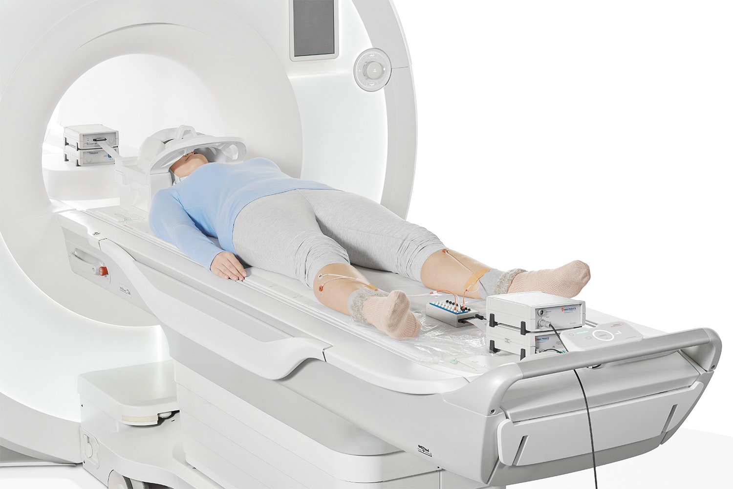
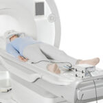
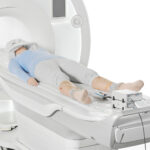
Exactly like all other amplifiers belonging to this product family, the BrainAmp ExG MR as well can be used inside the MRI chamber and placed right next to the subject. This allows to keep the length of the cables which are used as short as possible, thus ensuring highest data quality and fulfillment of all safety requirements.

The MR conditional auxiliary sensors (e.g. GSR MR Sensor, 3D Acceleration Sensor MR, Respiration Belt MR) offered by Brain Products are powered directly from the amplifier which guarantees patient safety and product portability.

BrainAmp ExG MR (16ch) can either be purchased as extension to the BrainAmp MR and BrainAmp MR plus, or as fully independent unit.

Technical Specifications

BrainAmp ExG MRTechnical specifications
Amplifier
Input impedance10 MΩ (for DC)
Filter typeFirst-order filter with 6 dB/octave
Lower cutoff frequency (high pass)0 Hz in DC mode or 0.016 Hz/10 s in AC mode; switchable between AC and DC modes
Filter typeFifth-order Butterworth filter with 30 dB/octave
High-cutoff frequency (low-pass)/time constant1000 Hz / 250 Hz (switchable for the resolutions 0.1 μV / 0.5 μV per bit)
Input noise≤ 2 μVpp
Common-mode rejection (CMR)≥ 100 dB
Bit width of the A/D converter (EEG and AUX)16 bit
Amplitude accuracy±2%
DC offset tolerance±300 mV
Integrated impedance measurementPresent; measurement includes ground electrode and reference electrode
Signal transmissionOptically coupled using duplex fiber optic cables
Power SupplyPowerPack (external rechargeable battery)
Power consumption (in operation)Max. 150 mA
Power consumption (in standby)Typically 7 mA
Operating time between chargesTypically 30 hours with one amplifier, 15 hours with two amplifiers
Dimensions (H x W x D)Approx. 68 mm x 160 mm x 187 mm
WeightApprox. 1.1 kg
Unlocking functionPresent
Locking of unused channelsPresent
Suitable for use in MR scanner roomYes

Related Products

The LOOP-IT's front panel is depicted with its touchscreen interface, 8x EEG inputs, 2x tES outputs, 4x BNC outputs, and a special port.
The neuroConn LOOP-IT is an all-in-one solution for closed loop EEG-tES, TMS-EEG, and EEG-tFUS.

neuroConn LOOP-IT

neuroConn DCMC is pictured with its control panel PC, keyboard, and mouse, along with 4x stimulation channels connected to a glass head via rubber electrodes.
An expandalbe multi-channel transcranial electrical stimulation (HD-tES) platform.

neuroConn DC-STIMULATOR MC

neuroConn DC-STIMULATOR PLUS is depicted laying on a table with a pair of rubber/sponge electrodes connected.
A capable and powerful one-channel transcranial electrical stimulation (tES) tool.

neuroConn DC-STIMULATOR PLUS

The BESA Connectivity software interface is a workflow-based GUI that walks you through the connectivity analysis of your choosing. Here you see the results of a Coherence analysis between multiple regional sources (source space, frequency anlaysis).
BESA Connectivity is a stand-alone module that extends BESA Research with streamlined, user-guided workflows for time–frequency and connectivity analysis of EEG and MEG data (in both scalp and source space). With multiple time–frequency and connectivity analysis methods supported, you can run batch analyses across subjects and easily visualize results using a variety of 2D and 3D renderings. The output is also ready for direct import into BESA Statistics.

BESA Connectivity 2.0

View Other Application Fields

Website Search

Schedule a call with our Consultants​

Please select your area to schedule an introductory call with one of our Scientific Consultants.

script here