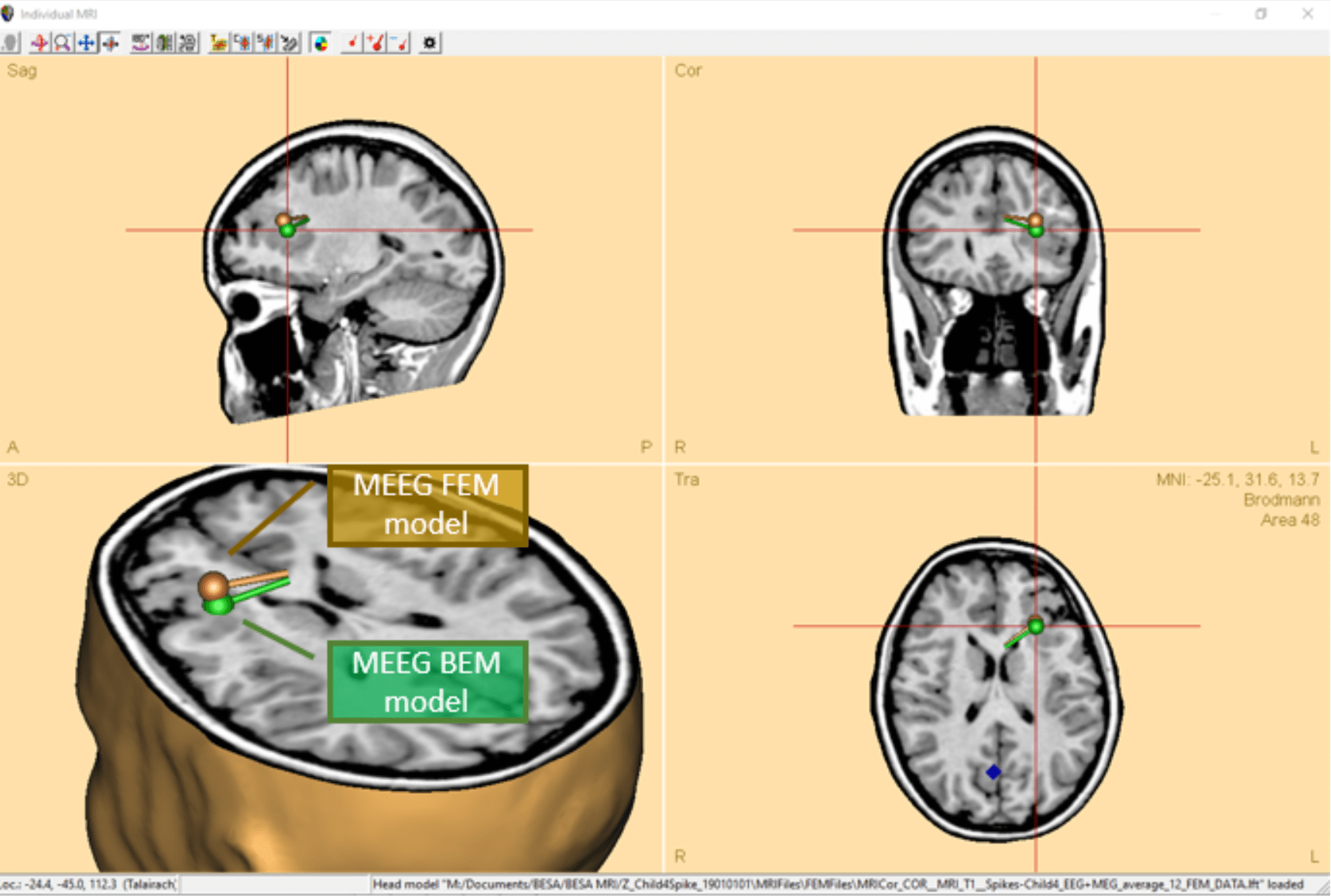

- For the first time, Boundary element method (BEM) and Finite element method (FEM) models can be directly compared on M/EEG data with just a couple of mouse clicks! After calculating the models automatically in BESA MRI, the models are automatically loaded into the Source analysis module and can be exchanged by two clicks in the head model section of the module.
- MEG and EEG can now be combined into a single data set for simultaneous source modeling of the two modalities. This is available for many source analysis and source imaging methods.
- MRI display now offers multi-slice view in any orientation for an easier review of solutions in the individual anatomy.
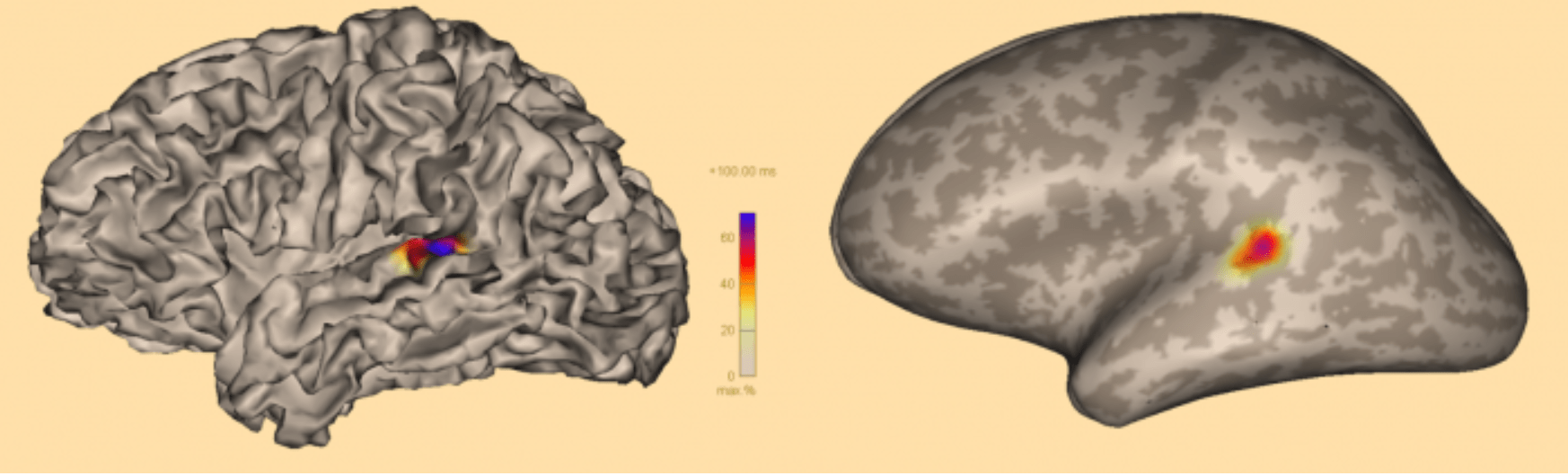
- Confidence limits are calculated for discrete source solutions, and displayed in co-registered MRI images.
- The Bayesian source imaging method SESAME was improved to enhance robustness, as well as speed of computation and convergence. For this purpose, hyper-priors were introduced (cf. https://arxiv.org/abs/2006.04141) and parallel computing was optimized for this method.
- The full noise covariance matrix computed from individual trials can now be used in computation of minimum norm estimates.
- Calculation of beamformer virtual sensor montages based on atlas regions is now supported.
Data review and pre-processing
- Atlas-based source montages: Pre-computed atlas-based source montages are now available from the menu entry Montage/Source/Atlas montages as well as under the Src button in the control ribbon.
- Parallel computing is used for speed-up of many time-consuming tasks.
- Smoother and faster plotting of waveforms eases review of high-density M/EEG data.
- New data readers for XDF and Neuroscan CURRY 8 formats are available.
Contact us for a free trial to test how BESA Research could fit into the data processing pipeline for your next research project!