Home › Products › Page 2
Filter
Vendors
Application Fields
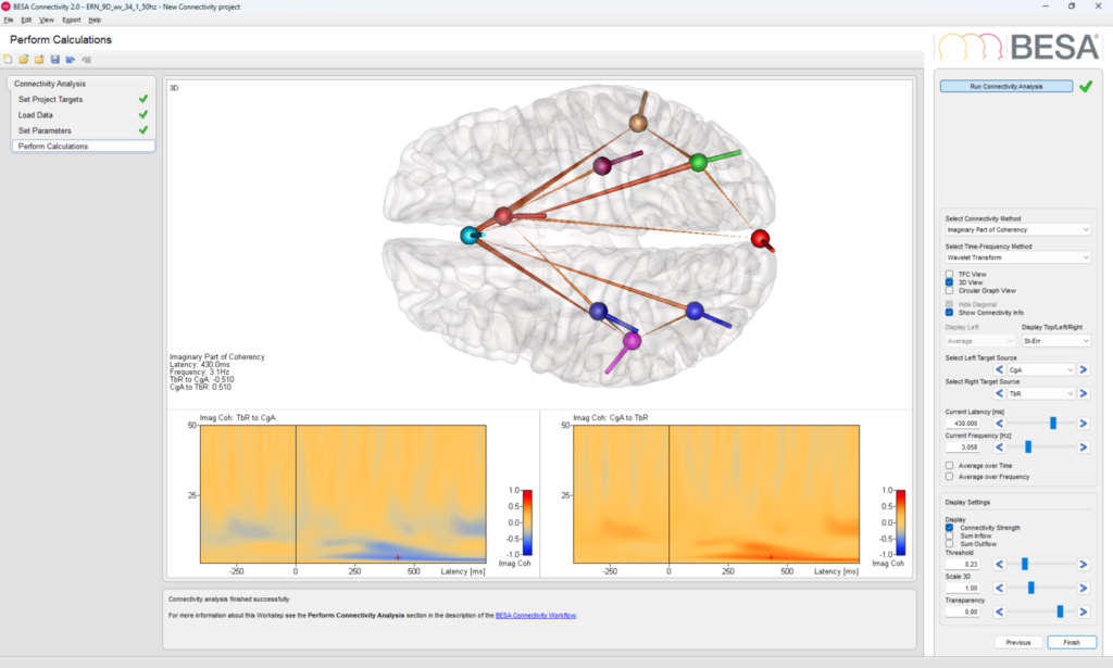
BESA Connectivity 2.0
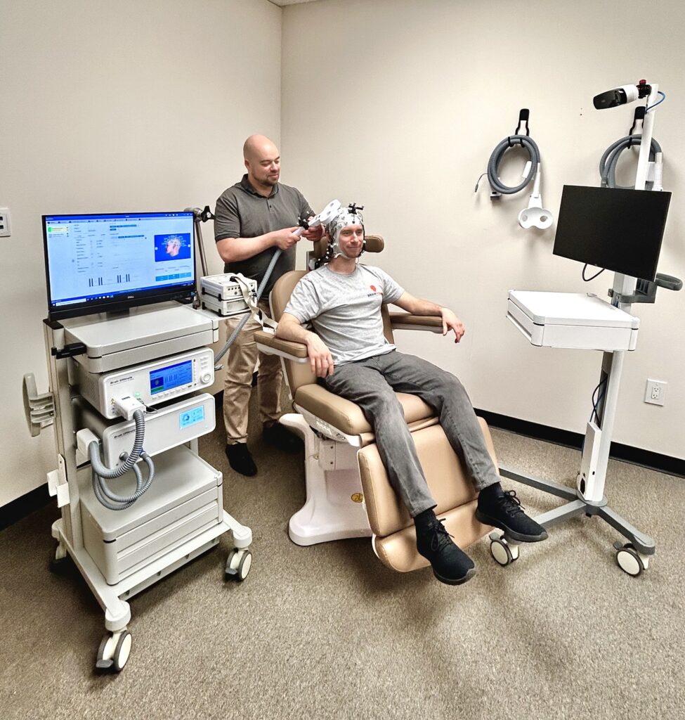
Brain Ultimate M-100 TMS System
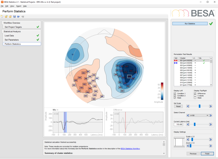
BESA Statistics 2.1
Tobii Pro Lab
Tobii Pro Spectrum
Tobii Pro Fusion
Tobii Pro Glasses
Brain Ultimate TMS-EEG Coils
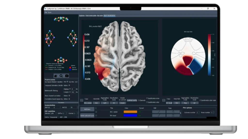
Cortivision CortiPrism
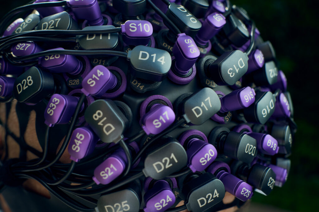
Cortivision SPECTRUM DOT
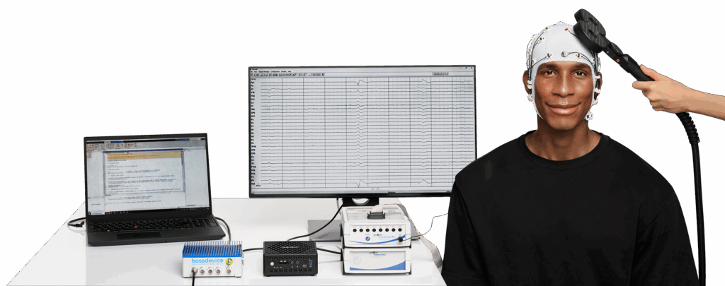
bossdevice RESEARCH with bossapp RESEARCH
Snaptrode (B18A) Bundles
Please select your area to schedule an introductory call with one of our Scientific Consultants.
script here