For more information or to speak with the
Brain Vision Scientific Consulting Team dial
(877) EEG-4MRI or schedule a call
Menu
Home » BESA MRI 3.0
Home » BESA MRI 3.0
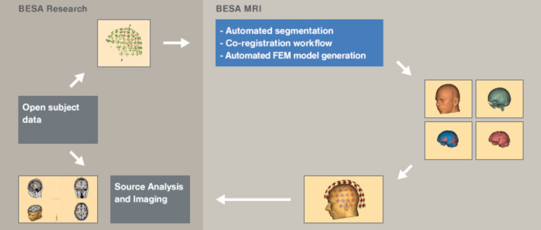
BESA MRI is software to generate individual head models (BEM / FEM) that can be used for EEG and / or MEG source analysis. BESA MRI also allows co-registration of EEG / MEG data with individual MRI data, and visualization of dipole solutions generated in BESA Research in the individual anatomy. BESA MRI is the first software that offers an easy, intuitive interface with an integrated workflow guiding the user step-by-step.
MRI data import is easily performed with DICOM, ANALYZE / NIFTI, VMR readers. The preparation of the MRI data to be automatically processed by BESA MRI requires just a couple of simple work steps typically demanding only a few minutes of user attention. BESA MRI’s automatic processing can be applied to all subjects prepared by the user in one go, therefore conveniently minimizing the time the user has to work on the computer.
BESA MRI’s automatic segmentation includes an automated inhomogeneity correction to correct for scan artifacts, generates a high quality cortex and scalp reconstruction with optional cortex inflation for enhanced visualization, and, in an optional step, can also generate an individual head model (BEM, FEM) with 3 layers (scalp, skull, brain – BEM) or 4 layers (scalp, skull, CSF and brain – FEM).
BESA MRI 3.0 runs in combination with BESA Research 7.1 and 7.0. Individual head models (BEM, FEM) generated with BESA MRI 3.0 can be imported by BESA Research 7.x for EEG and / or MEG source analysis. EEG / MEG data can be co-registered with individual MRI data using individually digitized electrode positions or head shape points. As a unique feature of BESA MRI 3.0, 10-10 standard electrodes (including inferior electrodes) can also be morphed with individual MRI data. Co-registered electrode coordinates are immediately available in BESA Research. Thus, source images / localizations can be displayed on the individual MRI even if no individual digitization of electrodes has taken place.
The MRI data can be visualized in a user-defined multi-slice view. Discrete solution files generated by BESA Research can be overlaid over the individual anatomy, as well as one of several available brain atlases.
For more information or to speak with the
Brain Vision Scientific Consulting Team dial
(877) EEG-4MRI or schedule a call
Billing address:
Brain Vision, LLC
1204 Village Market PL #300
Morrisville, NC 27560
Shipping address:
Brain Vision, LLC
515 N. Greenfield Parkway, Suite 100
Garner, NC 27529
| Cookie | Duration | Description |
|---|---|---|
| cookielawinfo-checkbox-analytics | 11 months | This cookie is set by GDPR Cookie Consent plugin. The cookie is used to store the user consent for the cookies in the category "Analytics". |
| cookielawinfo-checkbox-functional | 11 months | The cookie is set by GDPR cookie consent to record the user consent for the cookies in the category "Functional". |
| cookielawinfo-checkbox-necessary | 11 months | This cookie is set by GDPR Cookie Consent plugin. The cookies is used to store the user consent for the cookies in the category "Necessary". |
| cookielawinfo-checkbox-others | 11 months | This cookie is set by GDPR Cookie Consent plugin. The cookie is used to store the user consent for the cookies in the category "Other. |
| cookielawinfo-checkbox-performance | 11 months | This cookie is set by GDPR Cookie Consent plugin. The cookie is used to store the user consent for the cookies in the category "Performance". |
| viewed_cookie_policy | 11 months | The cookie is set by the GDPR Cookie Consent plugin and is used to store whether or not user has consented to the use of cookies. It does not store any personal data. |
* indicates required fields
"*" indicates required fields
Please select your area to schedule an introductory call with one of our Scientific Consultants.
script here